
This blog post article is part of a series of articles on assessment of the low back and pelvis. Scroll to the end of this article to see the others in this series.
Piriformis Stretch Test
The piriformis stretch test is used to assess piriformis syndrome, that is, compression of the sciatic nerve by the piriformis.
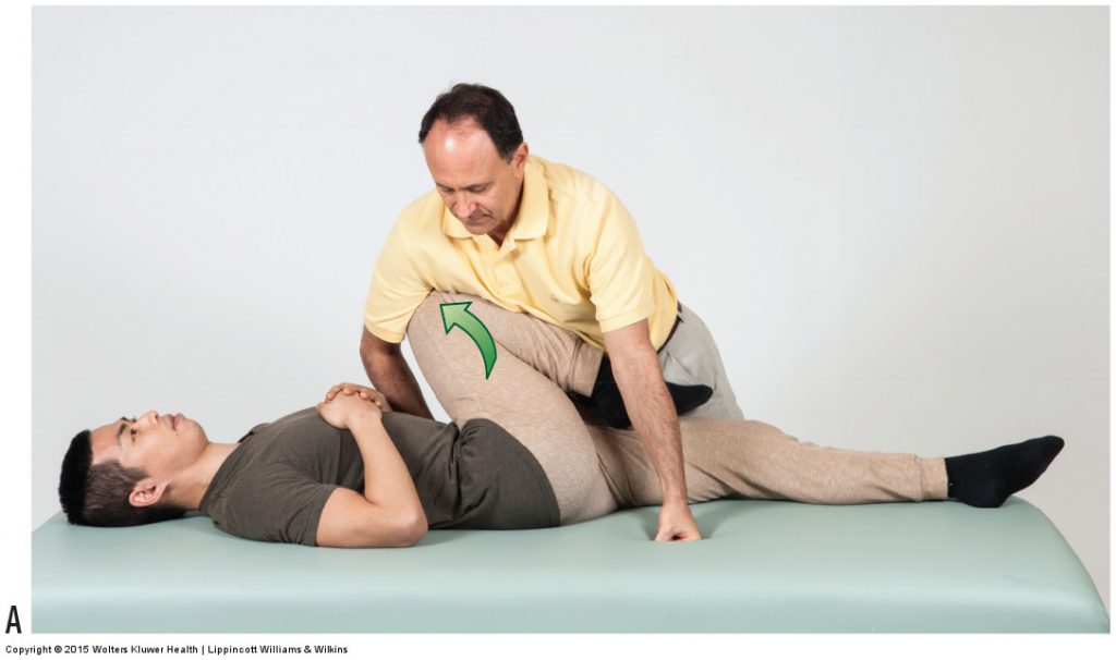
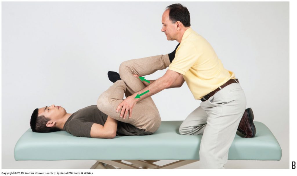
Figure 9. Stretches for the piriformis. (A) Horizontal adduction of the flexed thigh. (B) Lateral rotation of the flexed thigh. Permission Joseph E. Muscolino DC. Manual Therapy for the Low Back and Pelvis – A Clinical Orthopedic Approach (2015)
As its name implies, the piriformis muscle is stretched, thereby increasing its tautness and its compression on the sciatic nerve. Stretching a muscle is done by simply moving the client’s body into the opposite of its joint actions. Two excellent stretches for the piriformis are shown in Figure 9. In Figure 9A, the client’s thigh is flexed and then horizontally adducted (horizontally flexed) because the piriformis is a horizontal abductor (horizontal extensor) when the thigh is first flexed. In Figure 9B, the client’s thigh is flexed and laterally rotated because the piriformis is a medial rotator when the thigh is first flexed. If the client experiences a pulling (stretching) sensation or pain in the buttock, it is likely due to a tight piriformis but does not indicate compression of the sciatic nerve, therefore the test is not considered to be positive. For the piriformis stretch test to be considered positive, the client must experience referral of symptoms distally into the lower extremity (thigh, leg, and/or foot), indicating compression of the sciatic nerve by the piriformis muscle, in other words, piriformis syndrome.
Note: Piriformis and the Sciatic Nerve
The relationship between the piriformis muscle and sciatic nerve can vary. Usually, the sciatic nerve exits the pelvis inferior to the piriformis. However, some or all of the nerve may exit through or above the piriformis. Regardless of the relative relationship between these two structures, a tight piriformis can potentially compress the sciatic nerve and cause piriformis syndrome.
Note: Piriformis Syndrome and Sciatica
When the sciatic nerve is compressed by the piriformis muscle, it is often termed “pseudo-sciatica.” However, any compression upon the sciatic nerve is sciatica, regardless of whether the compression is a space-occupying lesion in the intervertebral foramen (IVF), usually caused by a pathologic disc or a bone spur, or by a tight piriformis muscle is technically sciatica.
(Click here for a blog post article on orthopedic assessment of thoracic outlet syndrome.)
This blog post article is the 12th in a series of 18 blog posts on the subject of assessment of the low back and pelvis.
The blog post articles in this series are:
- Introduction to Assessment of the Low Back and Pelvis
- Health History
- Introduction to Physical Assessment Examination of the Low Back and Pelvis
- Postural Assessment of the Low Back and Pelvis
- Range of Motion and Manual Resistance Assessment of the Low Back and Pelvis
- Muscle and Bone Palpation of the Low Back and Pelvis
- Joint Motion Palpation Assessment
- Overview of Special Orthopedic Assessment Tests of the Low Back and Pelvis
- Straight Leg Raise Tests for Space-Occupying Lesions
- Cough Test and Valsalva Maneuver
- Slump Test
- Piriformis Stretch Test
- Straight Leg Raise and Manual Resistance Tests for Strains and Sprains
- Nachlas and Yeoman’s Tests
- Sacroiliac Joint Medley of Tests
- Treatment Strategy for the Low Back and Pelvis
- Self-Care Advice for the Client with a Low Back / Sacro-Iliac Joint Condition
- Brief Review of Assessment and Treatment of Conditions of the Low Back and Pelvis
