- The sacroiliac joint (SIJ) is located between the sacrum and the iliac bone of the pelvic bone.
- The SIJ is a mixed synovial/fibrous joint.
- It allows small but important planar gliding motions of nutation and counternutation between the sacrum and ilium.
- Nutation is an anterior tilt movement of the sacral base relative to the ilium.
- Counternutation is an posterior tilt movement of the sacral base relative to the ilium.
NOTES:
- When we are born, the SIJ is a synovial diarthrotic joint; as we age and place continuing physical stress forces into the joint, it gradually evolves to become a fibrous amphiarthrotic joint.
- The SIJ is likely the most controversial joint in the human body. But even though its motions are small, they can be very important. And pain and dysfunction often occur at the SIJ.
- The posterior superior iliac spine (PSIS) is an extremely useful landmark when looking to palpate and locate the SIJ.
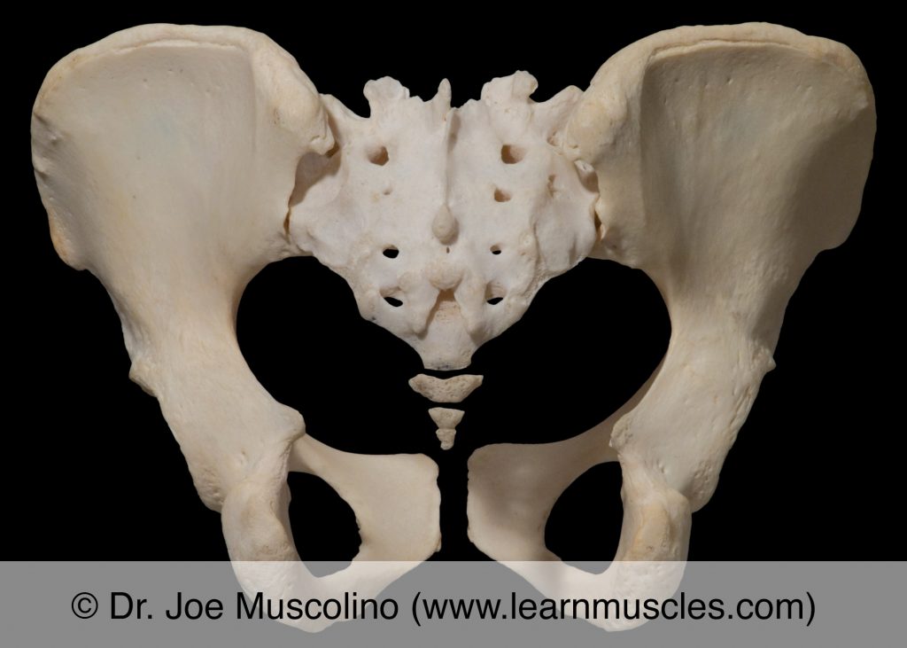
Posterior view of the sacroiliac joints (SIJs).
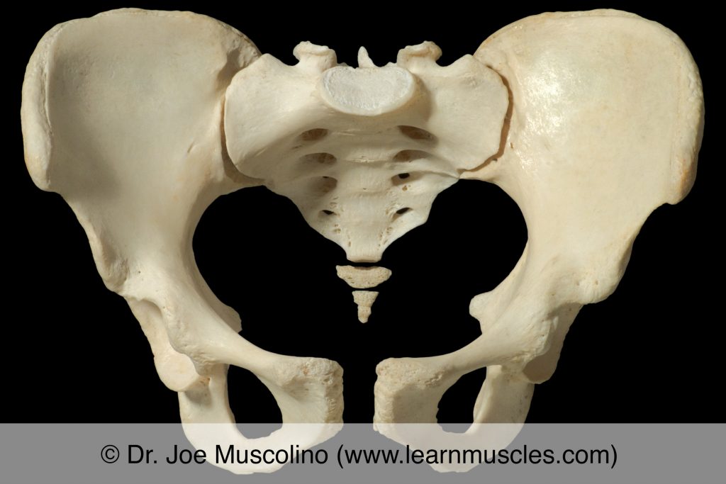
Anterior view of the sacroiliac joints (SIJs).
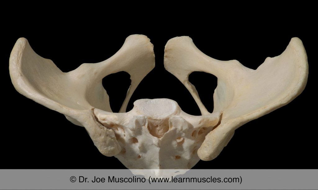
Superior view of the sacroiliac joints (SIJs).
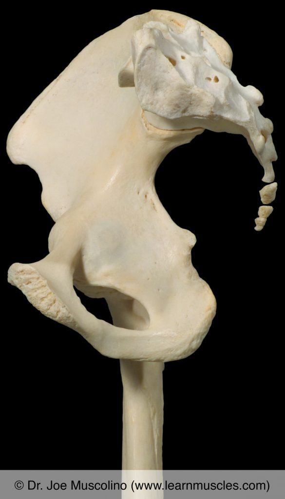
View from left to right of the sacrum articulating with the right-side pelvic bone. The articular surface of the sacrum on the left side is seen. The right femur is also seen.