This blog post article is part of a series of articles on assessment of the low back and pelvis. Scroll to the end of this article to see the others in this series.
Straight Leg Raise and Manual Resistance Tests
Following are assessment tests that are used to assess conditions caused by injury to soft tissues at a joint such as sprain, strain, and irritation/inflammation. Note: Even though straight leg raise (SLR) tests are forms of active and passive range of motion (ROM) tests and have therefore already been discussed under the “Range of Motion and Manual Resistance Assessment” section, they and manual resistance (MR) are discussed again here in their context to other assessment tests for injured tissue.
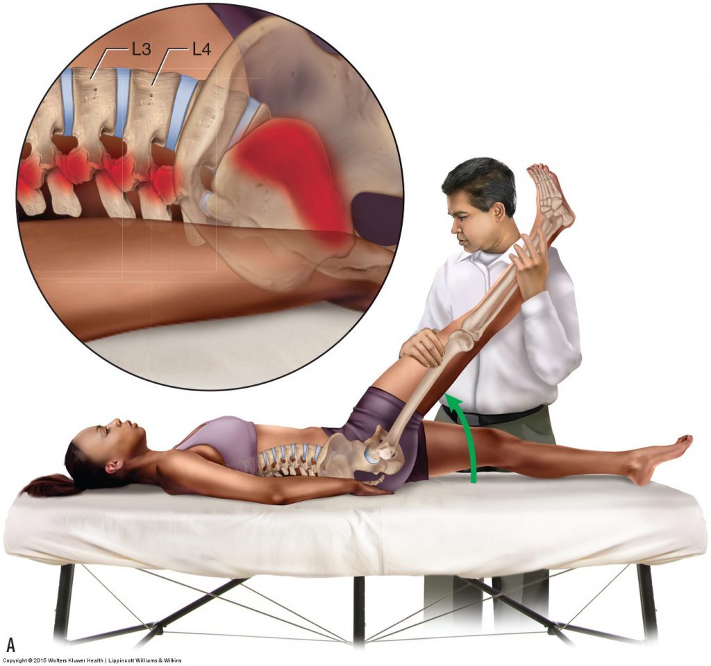
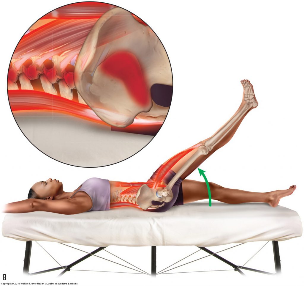
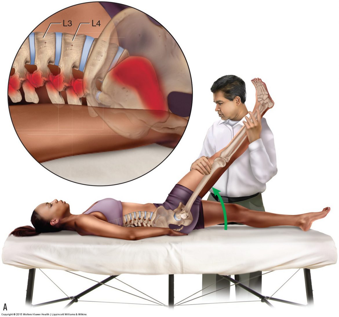
Figure 10. SLR tests to assess sprains and strains. (A) Passive SLR moves the sacroiliac and lumbar spinal joints and therefore assesses for a sprain of these joints. (B) Active SLR moves the joint tissues, and it requires musculature to engage; therefore, it assesses both sprains and strains.Permission Joseph E. Muscolino DC. Manual Therapy for the Low Back and Pelvis – A Clinical Orthopedic Approach (2015)
Passive Straight Leg Raise
Passive SLR test is performed as described earlier in this series of blog post articles in the section on special orthopedic assessment tests for space-occupying lesions. The difference is that now the intended assessment is for sprains and irritation/inflammation of the SIJ and lumbar spinal joints; the test is considered to be positive if local pain is felt in the SIJ region or lumbar spine (referral pain down the lower extremity is not the criterion for a positive test now). Passive SLR test is performed by the therapist raising the client’s thigh into flexion at the hip joint while keeping the client’s knee joint fully extended. It is important that the knee joint remains fully extended (hence the name straight leg raise) (Fig. 10A). Because the client is not engaging musculature of the region to create this motion, the client is passive. Flexing the thigh at the hip joint and keeping the knee joint fully extended places a stretch on the hamstrings. This pulls them taut, creating a tension/pulling force on the pelvic bone on that side. Therefore, as the client’s thigh is lifted into flexion, that side pelvic bone will be pulled into posterior tilt. Because the other pelvic bone is stabilized on the table and not moving, motion will occur between the pelvic bones at the SIJs. If either side SIJ is sprained/inflamed, pain will be felt locally at the SIJ.
Note: Straight Leg Raise and Sacroiliac Joint Assessment
When performing the SLR test to assess for sprains/inflammations and/or strains of the SIJ, although it is typical to assess the same-side SIJ as the thigh being raised, the SIJ on the opposite side of the body of the thigh being raised is also sometimes assessed. The mechanism for the SLR test is as follows: If the right thigh is flexed, then the right pelvic bone will move into posterior tilt. Because the left pelvic bone is stabilized on the table, it will not move. Therefore, motion occurs between the two pelvic bones. If the right SIJ is mobile, the right pelvic bone will move relative to the sacrum at the right SIJ. Therefore, motion is introduced into the right SIJ and if it is injured, pain would occur locally at that joint; therefore, the right SIJ is assessed. However, if the right SIJ is hypomobile and does not move, then when the right thigh is raised and the right pelvic bone posteriorly tilts, the sacrum will be “locked” to the right pelvic bone, and they will move as a unit relative to the left pelvic bone. This motion will occur at the left SIJ. When this occurs, if the left SIJ is injured, then pain would be felt at that joint and that joint is assessed. Further, even if the right SIJ is mobile and allows motion, as the thigh is lifted higher, there will be a point where the end of ROM of the right SIJ will be reached and motion will start to occur at the left SIJ, assessing the left-sided SIJ. For these reasons, regardless of which thigh is lifted into flexion, either SIJ could be assessed. Even though performing SLR test on one side could assess either SIJ, it is usual and customary to perform this test on both sides of the client’s body.
Most commonly, an SIJ sprain/inflammation will be felt when the thigh is at approximately 30 degrees of flexion. As the thigh is lifted higher than 30 degrees, the sacrum will posteriorly tilt with the pelvic bone, and as the sacrum reaches the end of its posterior tilt ROM at the lumbosacral joint relative to the L5 vertebra, it will begin to pull L5 into flexion. Once L5 is pulled into flexion to the end of its flexion ROM, L4 will flex. This motion will continue up to the lumbar spine as the thigh is moved farther and farther into flexion. If any of these lumbar spinal joints are sprained/inflamed, pain will be felt locally at that level. Generally, the higher the thigh is raised when pain occurs, the higher the region of the spine that is assessed.
Active Straight Leg Raise Test
Active SLR test is performed in a similar manner to passive SLR test except that the client is asked to actively raise the thigh into flexion at the hip joint by engaging the hip flexor muscles (Fig. 10B). This will also cause pelvic and lumbar musculature to engage to stabilize the pelvis and spine as the thigh is lifted. (Note: It will also cause the psoas major to contract as a mover of the thigh. When contracting, the psoas major will exert tension on its spinal attachments as well as the thigh, causing compression and anterior shear of the lumbar spine, which could add to the local low back pain of active SLR.) The joints are being moved in an identical manner as when the test was performed passively; therefore, active SLR test will assess sprains and irritations/inflammations of the sacroiliac and lumbar spinal joints just as passive SLR test did. However, because the client is actively contracting the musculature of the region, active SLR test will also assess strains of musculature. Active SLR test is considered to be an excellent screening test for the region because it will be positive with both sprains (and inflammations) of the joint ligaments and capsules, and strains of the musculature of the joint.
Note: Straight Leg Raise Test and Antagonist Musculature
It is usually stated that passive SLR test assesses only sprains, and that active SLR test assesses both sprains and strains. However, this is a bit of an oversimplification. Both passive and active SLR tests also assess for strains and spasms of the antagonistic musculature of the joint(s) because both tests require them to stretch to allow the joint to move (whether the joint moved passively or actively).
Manual Resistance Test
MR is another test that is used to assess strains of musculature. As its name implies, MR test is performed by the therapist using his or her hands to add resistance to the client’s attempted joint motion. The MR must be strong enough to stop the client from actually moving the joint. Therefore, the contraction is isometric. If the client’s musculature engages but no joint motion occurs, then the musculature will be stressed and assessed, but the joint ligaments and capsule will not. Therefore, MR test assesses for strain of the mover muscles of the joint, but not sprains of the joint.
This blog post article is the 13th in a series of 18 blog posts on the subject of assessment of the low back and pelvis.
The blog post articles in this series are:
- Introduction to Assessment of the Low Back and Pelvis
- Health History
- Introduction to Physical Assessment Examination of the Low Back and Pelvis
- Postural Assessment of the Low Back and Pelvis
- Range of Motion and Manual Resistance Assessment of the Low Back and Pelvis
- Muscle and Bone Palpation of the Low Back and Pelvis
- Joint Motion Palpation Assessment
- Overview of Special Orthopedic Assessment Tests of the Low Back and Pelvis
- Straight Leg Raise Tests for Space-Occupying Lesions
- Cough Test and Valsalva Maneuver
- Slump Test
- Piriformis Stretch Test
- Straight Leg Raise and Manual Resistance Tests for Strains and Sprains
- Nachlas and Yeoman’s Tests
- Sacroiliac Joint Medley of Tests
- Treatment Strategy for the Low Back and Pelvis
- Self-Care Advice for the Client with a Low Back / Sacro-Iliac Joint Condition
- Brief Review of Assessment and Treatment of Conditions of the Low Back and Pelvis
