Motions of the Joints of the Pelvis
This blog post article is an overview of the motions of the joints of the pelvis: the paired left and right sacroiliac joints and the symphysis pubis joint. For more complete coverage of the structure and function of the low back and pelvis, Kinesiology – The Skeletal System and Muscle Function, 3rd ed. (2017, Elsevier) should be consulted.
Pelvic motion can be considered in two ways.
- The pelvis can move as a unit relative to adjacent body parts.
- relative to the thigh at the hip joint
- relative to the trunk at the lumbosacral joint
- Motion can also occur within the pelvis; this is called intrapelvic motion.
Motion of the Pelvis as a Unit at the Hip Joint(s)
When the pelvis moves as a unit, it can move relative to both thighs at the hip joints or relative to one thigh at the hip joint on that side. These motions are anterior and posterior tilt in the sagittal plane, depression and elevation in the frontal plane (depression is also known as lateral tilt; elevation is also known as hip hiking), and left rotation and right rotation in the transverse plane. It is helpful to understand that motions of the pelvis at the hip joint are reverse actions of standard action motions of the thigh at the hip joint. In other words, the same functional group of muscles that moves the thigh at the hip joint when the pelvis is stabilized/fixed also moves the pelvis at the hip joint when the thigh is stabilized/fixed. The thigh moves at the hip joint when the distal end of the lower extremity kinematic chain is free to move, in other words, open chain kinematics; the pelvis tends to move at the hip joint when the distal end of the lower extremity kinematic chain is stabilized/fixed, in other words, closed chain kinematics. Specific pelvic reverse actions relative to the same-side thigh are as follows: pelvic anterior tilt is the reverse action of thigh flexion, pelvic posterior tilt is the reverse action of thigh extension, pelvic depression is the reverse action of thigh abduction, pelvic elevation is the reverse action of thigh adduction, pelvic contralateral rotation is the reverse action of thigh lateral rotation, and pelvic ipsilateral rotation is the reverse action of thigh medial rotation (Fig. 12 and Table 2).
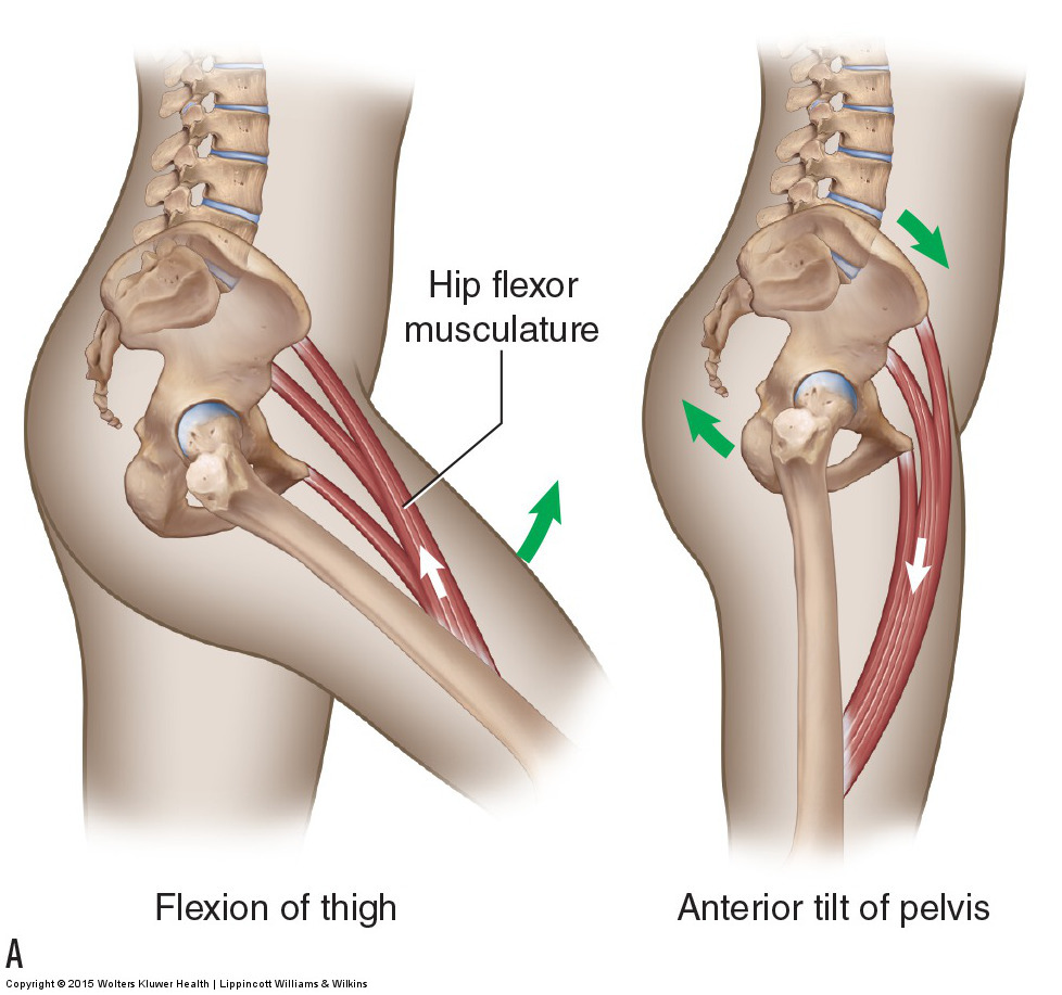
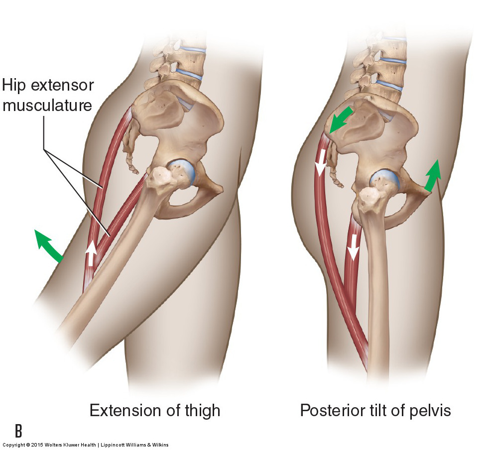
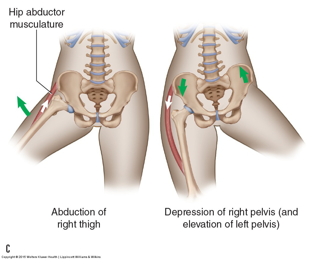
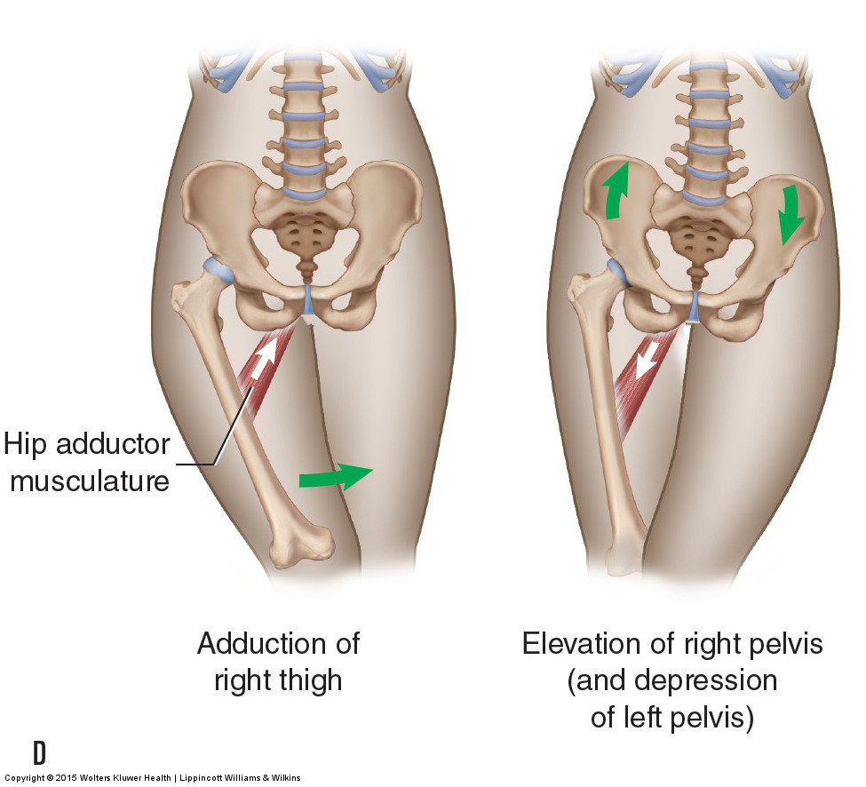
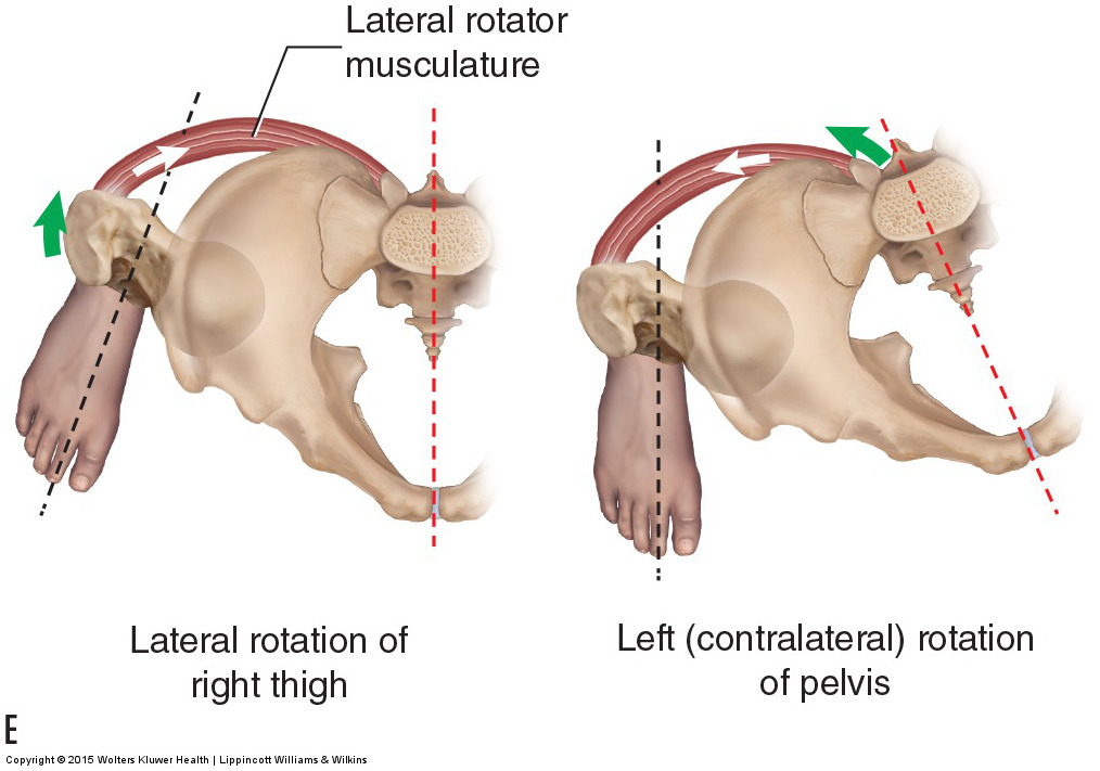

Figure 12. Joint actions of the pelvis at the hip joint are the reverse actions of the thigh moving at that hip joint. (A, B) Pelvic anterior tilt is the reverse action of thigh flexion; pelvic posterior tilt is the reverse action of thigh extension. (C, D) Pelvic depression is the reverse action of thigh abduction; pelvic elevation is the reverse action of thigh adduction. (E, F) Pelvic contralateral rotation is the reverse action of thigh lateral rotation; pelvic ipsilateral rotation is the reverse action of thigh medial rotation. Courtesy Joseph E. Muscolino. Manual Therapy for the Low Back and Pelvis – A Clinical Orthopedic Approach (2015).
Table 2. Standard (open chain) and Reverse (closed chain) Actions at the Hip Joint
| Standard Open Chain Thigh Actions | Reverse Closed Chain Pelvic Actions |
| Flexion | Anterior tilt |
| Extension | Posterior tilt |
| Abduction | Depression |
| Adduction | Elevation |
| Lateral rotation | Contralateral rotation |
| Medial rotation | Ipsilateral rotation |
Note: Horizontal Flexion and Extension
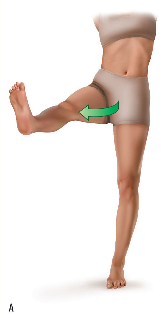
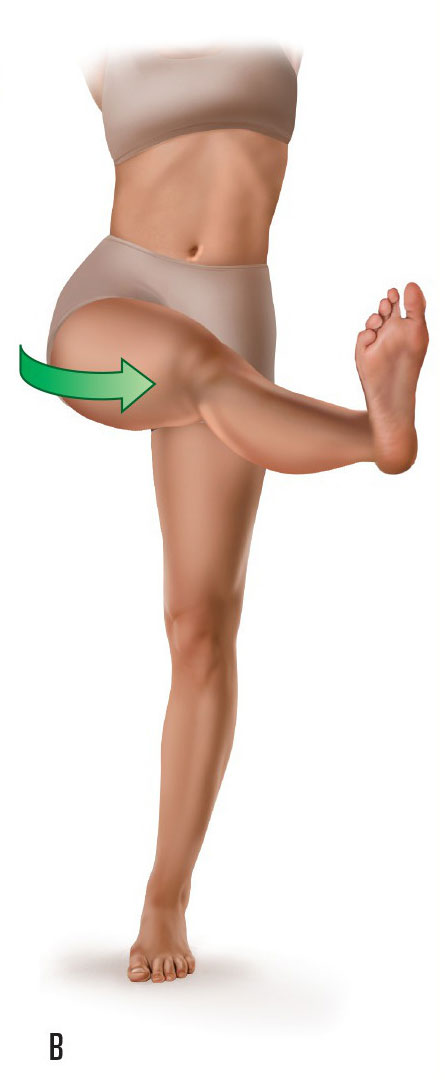
Two other motions of the thigh at the hip joint are named. They are horizontal flexion (also known as horizontal adduction) and horizontal extension (also known as horizontal abduction) (see accompanying figures). Horizontal abduction is a posterior/lateral motion of the thigh at the hip joint when the thigh is already flexed to 90 degrees (Fig. A); horizontal adduction is an anterior/medial motion of the thigh at the hip joint when the thigh is already flexed to 90 degrees (Fig. B). Horizontal adduction is useful when stretching the musculature of the posterior pelvis.
Motion of the Pelvis as a Unit Relative to the Lumbosacral Joint
The pelvis can also move relative to the trunk at the lumbosacral joint. These are the same named actions as when the pelvis moves at the hip joint: anterior and posterior tilt in the sagittal plane, elevation and depression in the frontal plane, and right rotation and left rotation in the transverse plane. Similar to pelvic motion being reverse actions of the thigh at the hip joint, pelvic actions can also be reverse actions of the trunk at the lumbosacral joint. When the inferior end of the body (the pelvis) is stabilized/fixed and the superior end of the body (the trunk) is free to move, the trunk moves at the lumbosacral joint (this could be looked at as open chain kinematics); when the superior end of the body (the trunk) is stabilized/fixed and the inferior end of the body (the pelvis) is free to move, the pelvis moves at the lumbosacral joint (this could be looked at as closed chain kinematics). Specifically, pelvic anterior tilt is the reverse action of trunk extension, pelvic posterior tilt is the reverse action of trunk flexion, elevation of the right side of the pelvis is the reverse action of trunk right lateral flexion, elevation of the left side of the pelvis is the reverse action of trunk left lateral flexion, pelvic rotation to the right is the reverse action of trunk rotation to the left, and pelvic rotation to the left is the reverse action of trunk rotation to the right (Fig. 13 and Table 3).
Table 3. Standard (open chain) and Reverse (closed chain) Actions at the Lumbosacral Joint
| Standard Open Chain Trunk Actions | Reverse Closed Chain Pelvis Actions |
| Extension | Anterior tilt |
| Flexion | Posterior tilt |
| Right lateral flexion | Right side elevation* |
| Left lateral flexion | Left side elevation* |
| Left rotation | Right rotation |
| Right rotation | Left rotation |
*When one side of the pelvis elevates, the other side of the pelvis depresses.
Note: Pelvic Posture and the Spine
The base (superior surface) of the sacrum forms the base upon which the spine sits. Therefore, if the posture of the pelvis changes, the posture of the sacral base changes, and the posture of the spine changes. For this reason, pelvic posture is critically important to the posture of the spine, and it is important for the therapist to be aware of all muscular, ligamentous, and other fascial structures that can affect the posture of the pelvis.
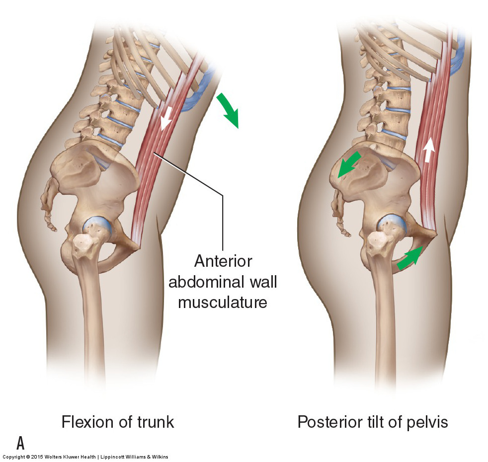
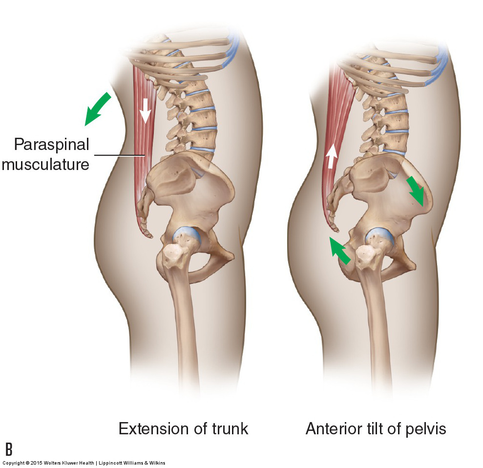
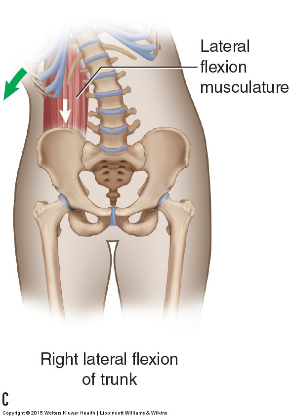
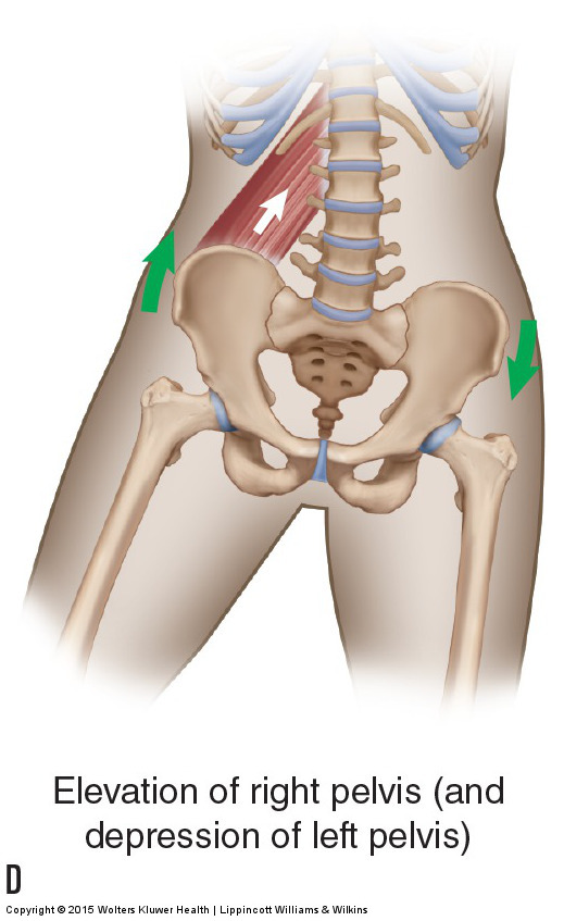
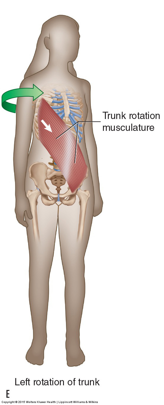
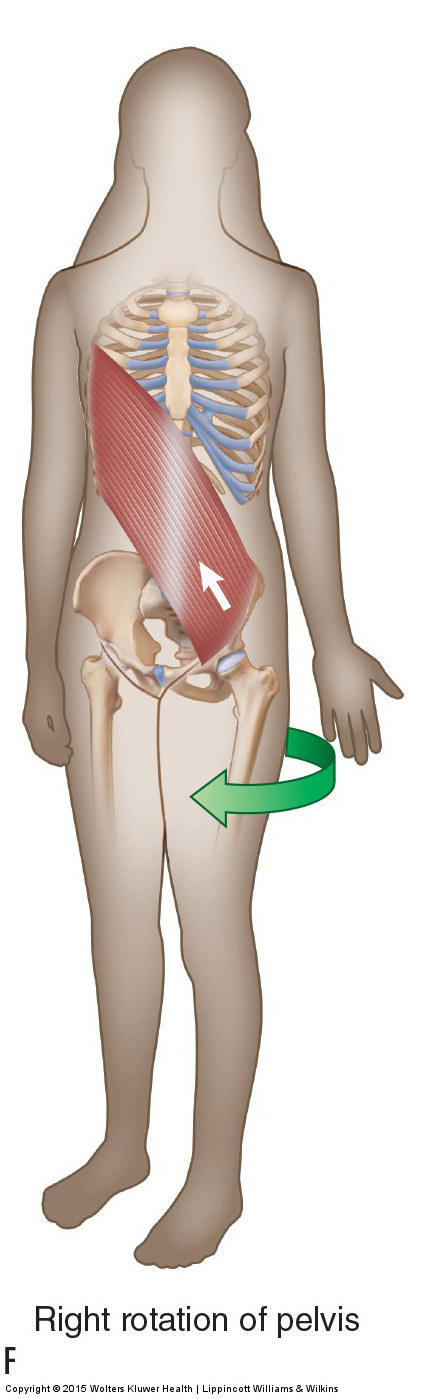
Figure 13. Joint actions of the pelvis at the lumbosacral joint are the reverse actions of the trunk moving at the lumbosacral joint. (A, B) Pelvic posterior tilt is the reverse action of trunk flexion; pelvic anterior tilt is the reverse action of trunk extension. (C, D) Pelvic right side elevation is the reverse action of trunk right lateral flexion (pelvic left side elevation is the reverse action of trunk left lateral flexion). (E, F) Pelvic right rotation is the reverse action of trunk left rotation (pelvic left rotation is the reverse action of trunk right rotation). Courtesy Joseph E. Muscolino. Manual Therapy for the Low Back and Pelvis – A Clinical Orthopedic Approach (2015).
Even though the pelvis can move at the hip joint(s) and the lumbosacral joint, pelvic motion at the hip joints is functionally more important, both from the standpoint of posture and motion.
Intrapelvic Motion
Motion can also occur within the pelvis; this is termed as intrapelvic motion. Intrapelvic motion involves motion of a pelvic bone relative to the sacrum or motion of the sacrum relative to the pelvic bone at the sacroiliac joint located between them (this also involves motion between the pelvic bones at the symphysis pubis joint anteriorly). These motions can be named for the sacral motion that occurs or for the motion of the pelvic bone.
When describing the sacral motion within the sagittal plane (or near sagittal plane), the terms nutation and counternutation are used. When the sacral base drops anteriorly, it is called nutation; when the sacral base moves in the posterior direction, it is called counternutation (Fig. 14). Nutation can also be described as anterior tilt; counternutation can also be described as posterior tilt.
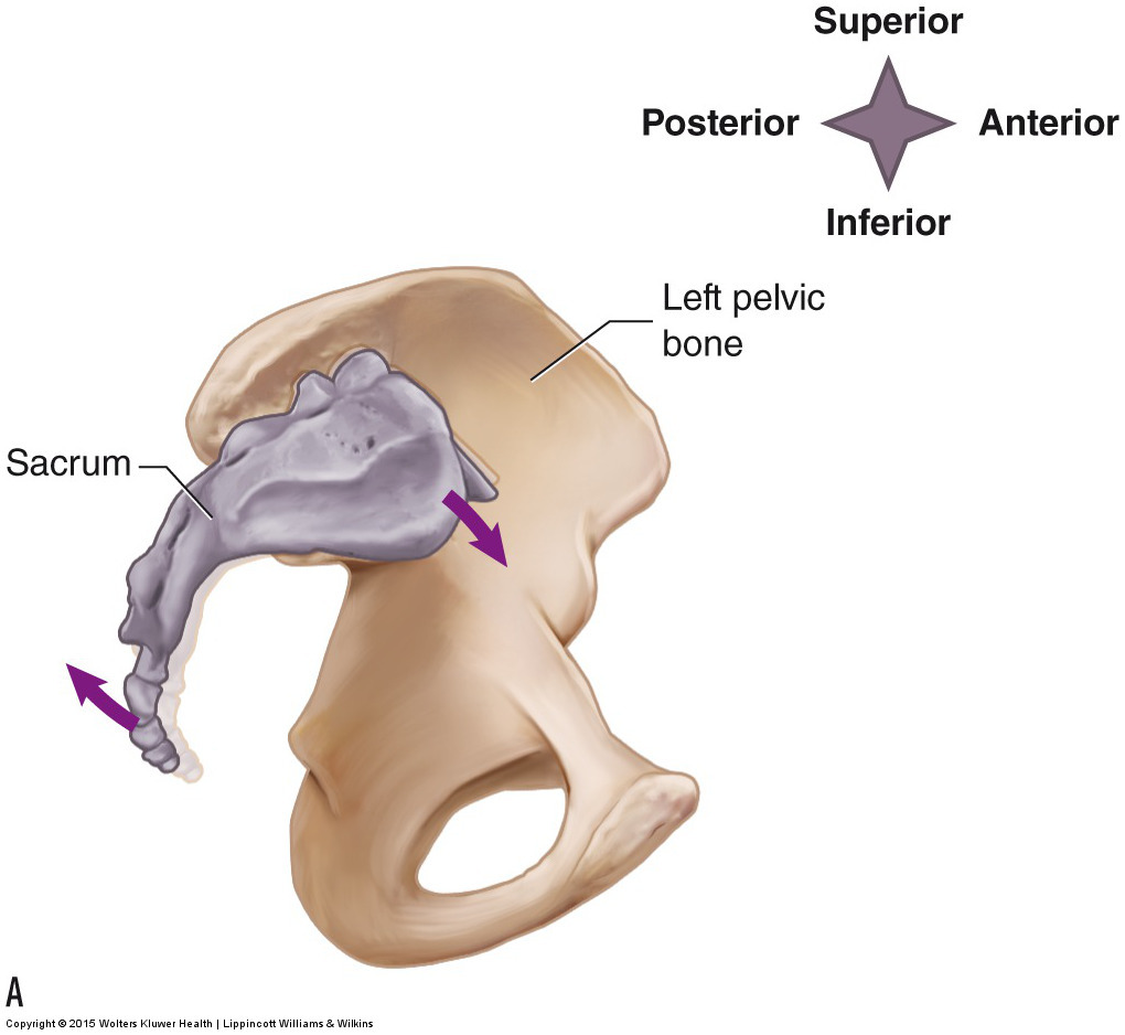

Figure 14. Right lateral view of nutation and counternutation of the sacrum. (A) Nutation. (B) Counternutation. Courtesy Joseph E. Muscolino. Manual Therapy for the Low Back and Pelvis – A Clinical Orthopedic Approach (2015).
When describing motion of the pelvic bone at the sacroiliac joint in the sagittal plane (or near sagittal plane), the terms posterior tilt and anterior tilt are used. In effect, posterior tilt of the pelvic bone is the reverse action of nutation of the sacrum at the sacroiliac joint, and anterior tilt of the pelvic bone is the reverse action of counternutation of the sacrum at the sacroiliac joint.
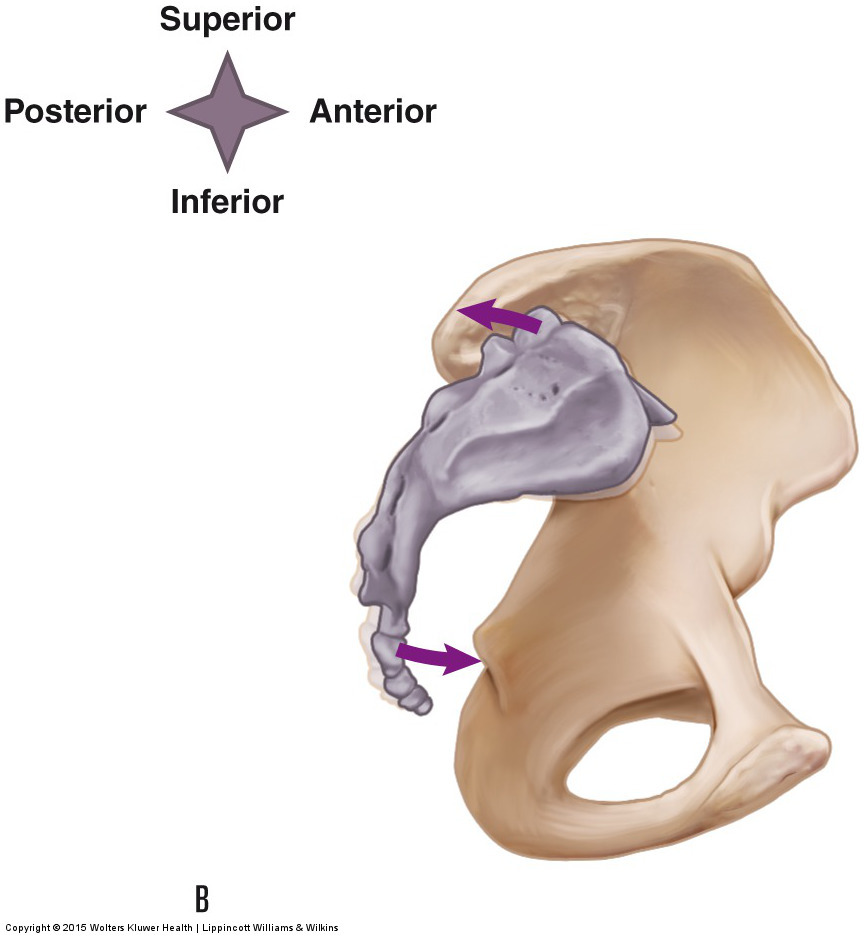
The pelvic bone can also be described as moving in the transverse plane. The term internal (or medial) rotation describes the anterior surface of the pelvic bone orienting medially (in effect, toward the opposite side of the body). This motion gaps (opens) the posterior aspect of the sacroiliac joint (and approximates [closes] the anterior aspect of the sacroiliac joint). The term external (or lateral) rotation describes the anterior surface of the pelvic bone orienting more laterally (toward the same side of the body). This motion gaps the anterior aspect of the sacroiliac joint (and approximates the posterior aspect of the sacroiliac joint) (Fig. 15). In effect, intrapelvic motion involves one side of the pelvis moving relative to the other side. Sacroiliac joint motions are small but are very important. An understanding of this motion is critically important when performing joint mobilization technique.
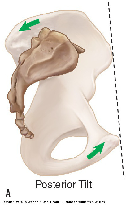
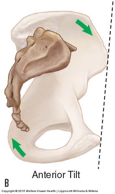
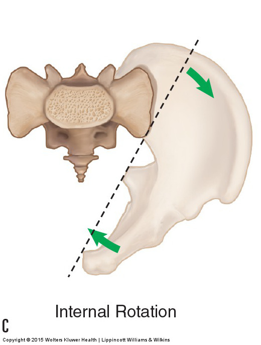
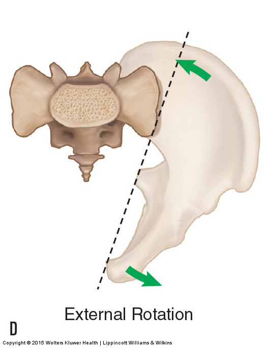
Figure 15. Motion of the pelvic bone at the sacroiliac joint. (A, B) Right lateral view of posterior tilt and anterior tilt, respectively. (C, D) Superior view of internal rotation and external rotation of the pelvic bone, respectively. Courtesy Joseph E. Muscolino. Manual Therapy for the Low Back and Pelvis – A Clinical Orthopedic Approach (2015).
Note: This is the fourth in a series of 8 blog post articles on the anatomy and physiology of the lumbar spine and pelvis.
The blog post articles in this series are:
- Bones of the Lumbar Spine and Pelvis
- Joints of the Lumbar Spine (disc & facet) and Pelvis
- Motions of the Joints of the Lumbar Spine
- Motions of the Joints of the Pelvis
- Muscles of the Lumbar Spine
- Muscles of the Pelvis
- Ligaments of the Lumbar Spine and Pelvis
- Precautions for Manual Therapy of the Lumbar Spine and Pelvis
