- The subtalar joint is located under the talus, hence its name subtalar joint.
- The subtalar joint is formed by the articulation of the talus with the calcaneus, therefore it is also known as the talocalcaneal joint.
- The subtalar joint is a synovial, diarthrotic, uniaxial joint.
- It allows:
- Pronation and Supination, which are oblique plane motions composed of:
- Pronation: eversion (frontal plane), abduction (transverse plane), and dorsiflexion (sagittal plane).
- Supination: inversion (frontal plane), adduction (transverse plane), and plantarflexion (sagittal plane).
NOTES:
- The subtalar joint is sometimes referred to as the lower ankle joint.
- The subtalar joint is uniaxial but triplanar because its oblique-plane uniaxial movements of pronation and supination are across all three cardinal planes.
- Frontal-plane eversion is NOT the same as pronation; it is the principal cardinal-plane component of the oblique-plane motion of pronation.
- Similarly, frontal-plane inversion is NOT the same as supination; it is the principal cardinal-plane component of the oblique-plane motion of supination.
- Abduction of the foot can be described as lateral rotation; adduction of the foot can be described as medial rotation.
- Many people have a condition known as overpronation, also known as hyperpronation, or described as dropped arches or flat foot (supple flat foot or rigid flat foot).
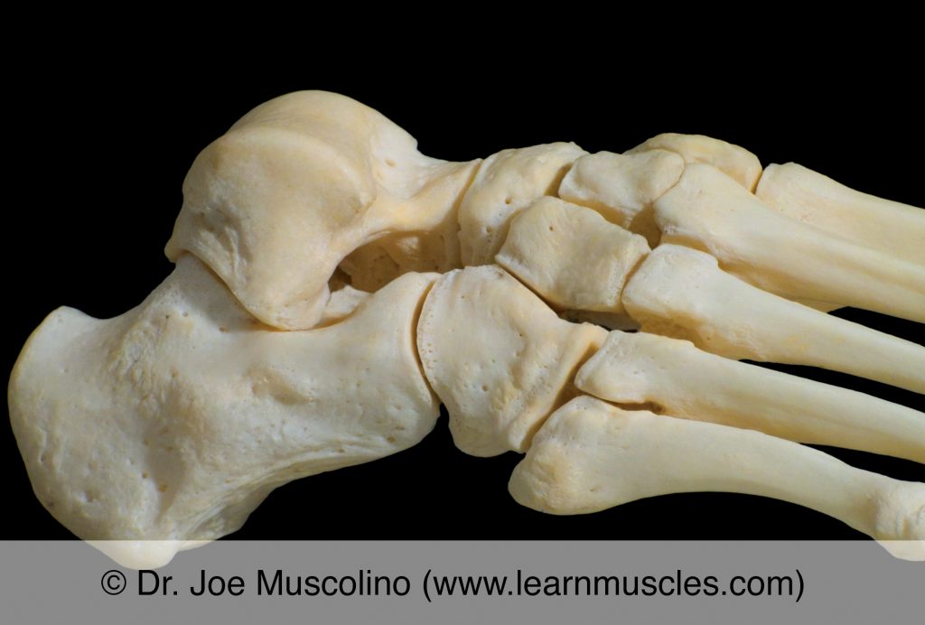
Lateral view of the subtalar joint on the right side of the body.
Lateral view of the right foot with the talus lifted away to better visualize the subtalar joint.
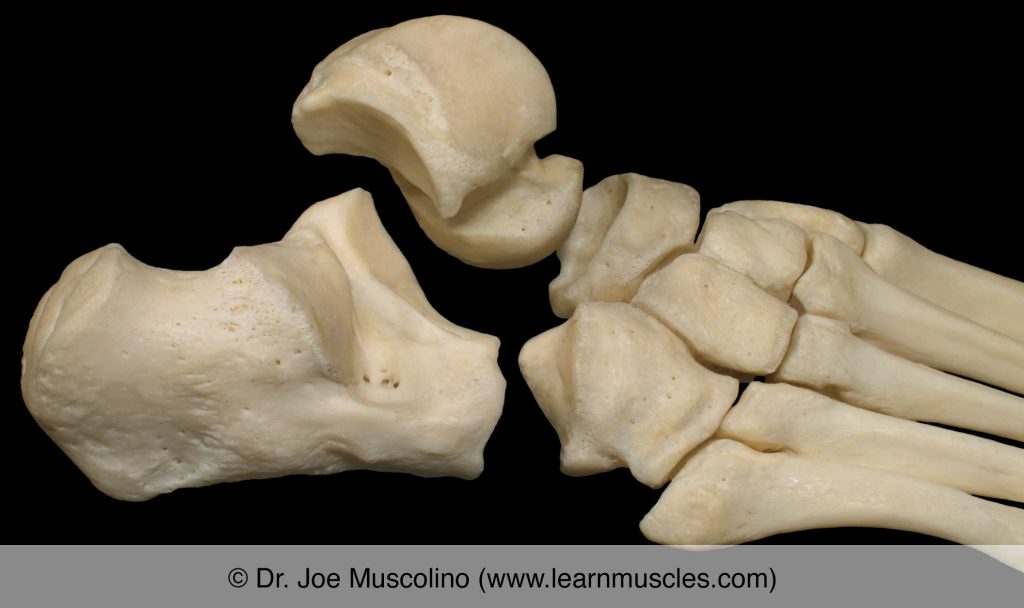
Talus and calcaneus of the right-side subtalar joint in open book view.

Medial view of the subtalar joint on the right side of the body.