- Typically, there are three spinal joints located between each two contiguous vertebrae: paired facet joints and a disc joint.
- the facet joints are posterior and lateral.
- the disc joint is anterior and midline.
- Facet joints are synovial, diarthrotic, planar joints that allow motion in all three cardinal planes.
- Disc joints are cartilaginous amphiarthrotic joints that allow motion in all three cardinal planes.
- Generally, facet joints determine the type of motion best permitted at that segmental joint level and the disc joint determines how much motion is possible.
- Spinal joints allow:
- Flexion/Extension in the sagittal plane.
- Right lateral flexion / Left lateral flexion in the frontal plane.
- Right rotation / Left rotation in the transverse plane.
NOTES:
- Spinal joints are generally name for their segmental joint level. For example, T4-T5 is the joint level between T4 and T5, and L1-L2 is the joint level between L1 and L2.
- The joint between the lumbar spine and the sacrum, in other words between L5 and the base of the sacrum, is called the lumbosacral joint or the L5-S1 joint.
- Facet joints are technically named apophyseal joint (singular) and zygapophyseal joints (plural).
- Disc joints are technically named intervertebral disc joints.
- Facet joints are located between the inferior articular facets of the superior vertebra and the superior articular facets of the inferior vertebra.
- Disc joints are located between vertebral bodies.
- Facet joints determine the type of motion at their level based on the plane of their facet surfaces.
- Generally, facet joints in the cervical spine best allow transverse plane rotation; facet joints in the thoracic spine would best allow lateral flexion except that the rib cage inhibits much frontal plane lateral flexion; the facet joints in the lumbar spine best allow sagittal plane flexion/extension.
- The thicker the disc, the more motion that is allowed at that segmental joint level.
- Disc joints bear more weight than facet joints.
- The atlas (C1) has no body, so there is no disc joint between the atlas and occiput (atlanto-occipital joint) and no disc joint between the atlas and the axis (C2) (atlantoaxial joint). The atlanto-occipital joint has its own glossary post.
- The atlantoaxial (C1-C2) joint complex does have the atlanto-odontoid joint between the anterior arch of the atlas (C1) and the odontoid process (dens) of the axis (C2). The atlanto-axial joint has its own glossary post.
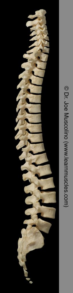
Right lateral view of the spinal column.
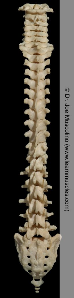
Anterior view of the spinal column.
Right lateral view of the cervical spine.
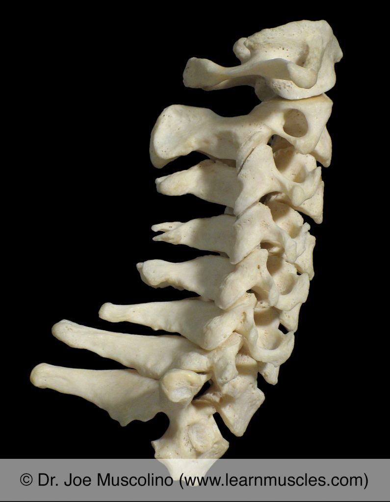
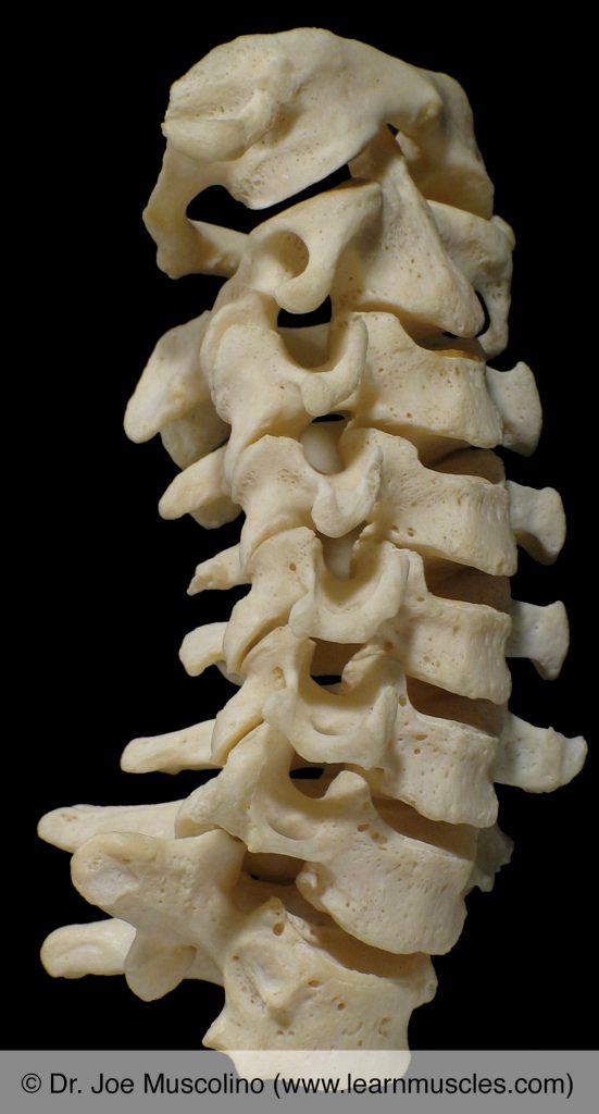
Right lateral view of the thoracic spine.
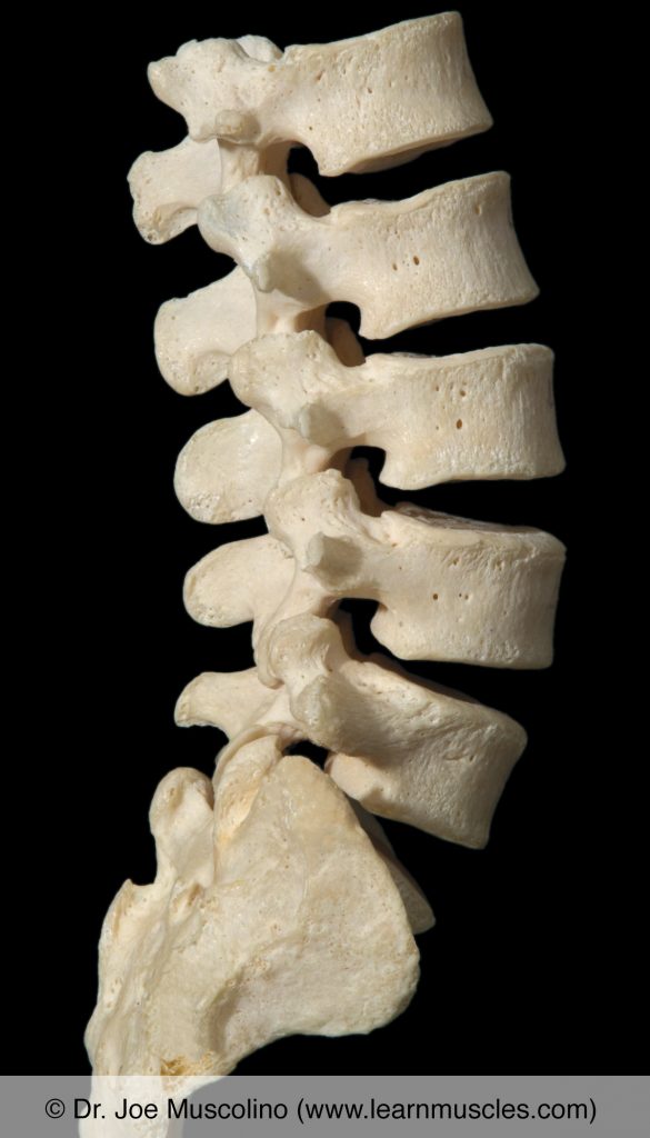
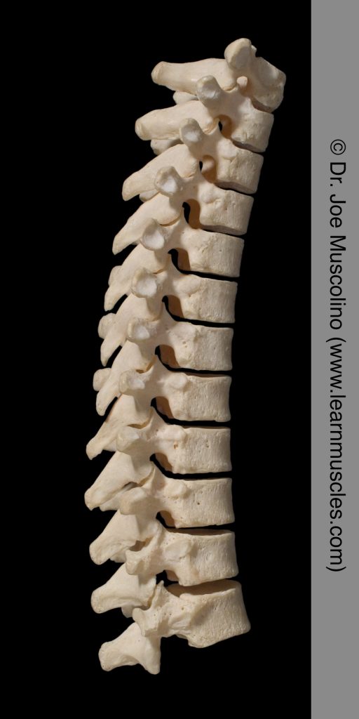
Right lateral view of the lumbar spine.
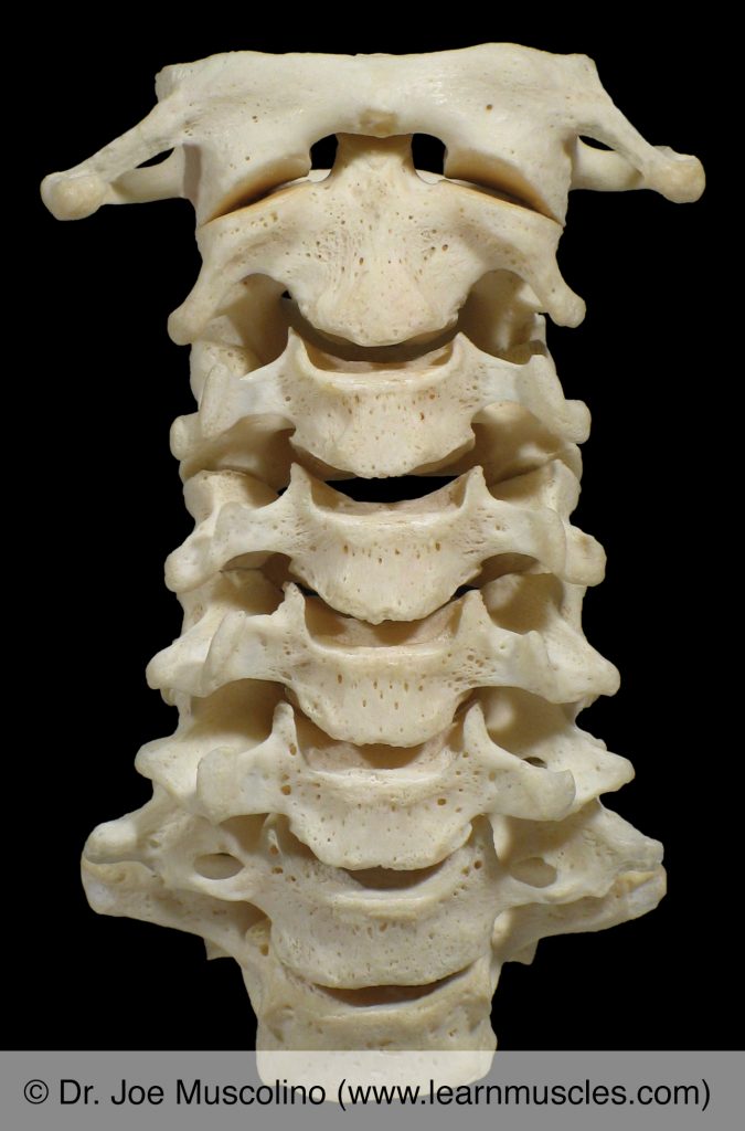
Anterior view of the cervical spine.
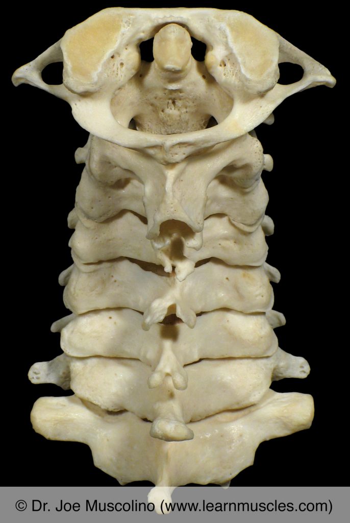
Posterior view of the cervical spine.
Posterior view of the thoracic spine.
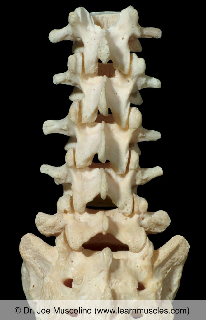
Posterior view of the lumbar spine.
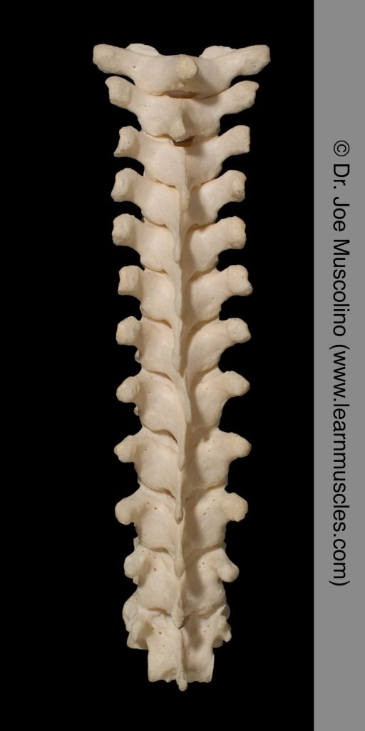
Oblique (anterolateral) view of the cervical spine.