
Signs and symptoms of a low back sprain and strain:
A low back sprain and strain present a similar clinical picture. The client/patient will have low back pain and spasming, and these symptoms will increase with motion. Most problematic is flexion (bending over) because this places a tension force on the torn posterior tissue(s). If the strain/sprain is focused on one side of the low back, lateral flexion to the opposite side will also increase the pain and spasming. However, because of the protective muscular spasming that occurs with a low back strain/sprain, motion in any direction can be limited. Pain that accompanies motion will also tend to decrease the client/patient’s low back range of motion. Acute strains and sprains also present with inflammation; generally the degree of inflammation reflects the degree of tearing.
Assessment/Diagnosis of a low back sprain and/or strain:
When assessing a low back sprain and/or strain, the verbal history is important. If a macrotrauma occurred, whether it was a strain or a sprain, the client/patient will often recall that they felt a “pop” or something “give” when they were bending over; and that the onset of pain was immediate.
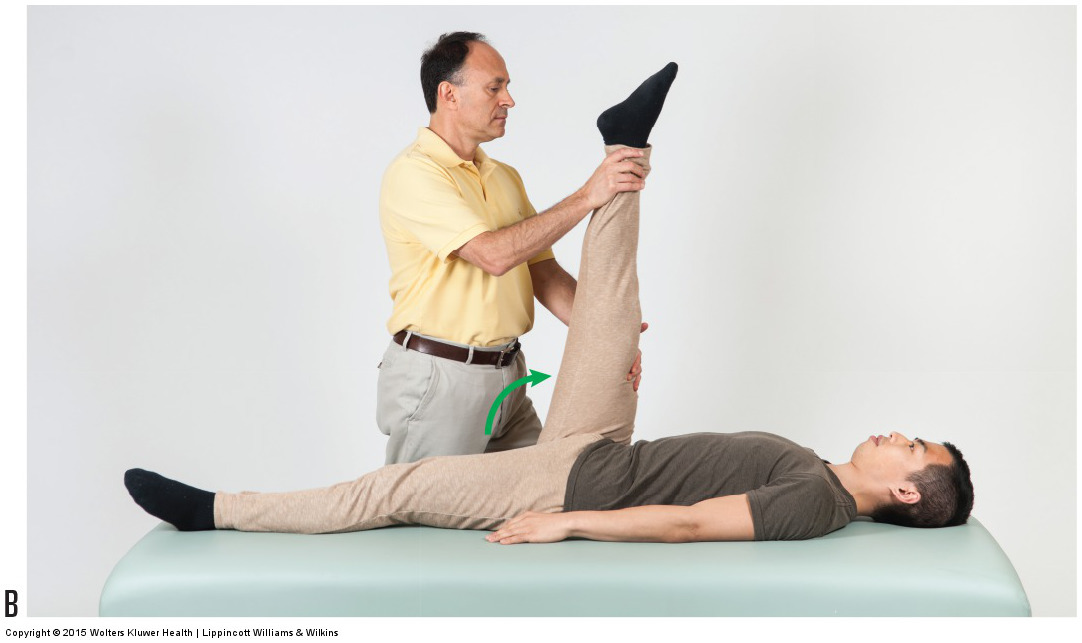
Passive straight leg raise orthopedic test. Permission: Joseph E. Muscolino. Manual Therapy for the Low Back and Pelvis – A Clinical Orthopedic Approach (2015).
Physical examination assessment of a low back sprain / strain is carried out using active and passive straight leg raise (SLR) as well as manual resistance (MR); in each case, local low back pain is the criterion for the test being positive. Active SLR will show positive for either a strain or a sprain. Active SLR assesses a strain because muscular tissue contracts to stabilize the spine during the motion; active SLR assesses a sprain because fascial ligamentous tissue is stretched when the joints are moved. Therefore, if either tissue is torn, this stress will evoke pain.
Passive SLR will usually show positive only for a low back sprain because muscles do not need to contract, but the joints are still moved so the ligaments are stretched. (Note: It is possible for passive SLR to cause pain with a muscular strain because passive SLR does require antagonistic tissue to stretch, thereby causing pain if strained. if the paraspinal muscle tissue is tight enough, then stretching it with passive SLR can possibly cause pain). If desired, MR can also be used in the assessment process. Ask the client/patient to attempt to perform active SLR but do not let them move the leg. This will engage musculature but will not move the joints, so MR should be positive only with a strain (Table 1).
Table 1: Assessment flow chart for a low back strain/sprain.
- Active SLR – Positive with low back sprain and/or strain
- Passive SLR – Positive with low back sprain only (may be positive if paraspinal musculature is very tight)
- MR – Positive with low back strain only
*SLR = Straight Leg Raise
*MR = Manual Resistance
In addition to these tests simply being positive or negative for pain, it should also be noted where the pain is located, how intense it is, and at what point during the SLR motion it occurred. The location of the pain indicates the location of the injury. At what point during the SLR motion the pain occurred also gives an indication of where the injury is located. Sacroiliac joint injury (SIJ) will generally manifest when the thigh is flexed approximately 30 degrees; lumbosacral joint (LSJ) injury will manifest at approximately 40 degrees; and the higher the thigh is flexed when the pain occurs, generally the higher in the lumbar spine the injury is located. How intense the pain is gives an indication of how severe the injury is.
The next step when assessing a low back sprain and/or strain is to palpate for the injured low back tissue(s). If a strain was assessed, each of the muscles of the low back should be palpated to determine which muscles are affected. Although low back strains usually involve the paraspinal muscles, it is important to also palpate and assess the other muscles of the low back, including the quadratus lumborum and psoas major.
If a low back sprain was assessed and the SIJ is suspected, palpation directly over the joint will usually be tender for the client/patient. Assessment of SIJ strains and sprains should include palpation assessment of the gluteal and piriformis muscles, as well as the coccygeus and levator ani muscles. If a SIJ or LSJ sprain is suspected, two further tests can be performed: Nachlas test and Yeoman’s test. In each case, pain at the joint is the criterion for the test to be positive.
Nachlas test is performed by bringing the foot of the prone client/patient toward the buttock on that side. This stretches the rectus femoris of the quadriceps group, placing an anterior tilt pulling force on the same-side pelvic bone. Because the other pelvic bone is stabilized with body weight, this will introduce motion into the same-side SIJ. If it is sprained, pain will occur. Nachlas test is then performed on the other side of the client/patient’s body. Nachlas can also assess the opposite-side SIJ as well as the LSJ joint. If the same-side SIJ is locked (hypomobile), then when the patient’s/client’s leg is moved, the anterior tilt force on that side pelvic bone will be transferred to the sacrum and the sacrum will move into anterior tilt (nutation). Because the opposite-side pelvic bone and the L5 vertebra are stabilized with body weight, this will place a force into both the opposite-side SIJ and the LSJ. Therefore, pain at either of these joints is positive for a sprain at that joint. Note: Nachlas test is contraindicated if the client/patient has a bad knee joint.
Yeoman’s test is performed by lifting the patient’s/client’s thigh into extension as pressure is placed on the same-side PSIS (to both stabilize the spine and also to add anterior tilt force to that pelvic bone). The biomechanics of Yeoman’s test are essentially the same as Nachlas test; only now all the hip flexors are stretched (not only the rectus femoris), placing a greater anterior tilt force on the same-side SIJ (as well as the opposite-side SIJ and LSJ). Because Yeoman’s test is more powerful, it is more sensitive at detecting a SIJ or LSJ sprain.
Differential diagnosis/assessment:
Both a low back sprain and low back strain involve protective muscular spasming, as does most every other condition of the low back. Therefore strains and sprains need to be differentially diagnosed/assessed from these other conditions. The first differential assessment to be made is to determine if the client/patient is simply experiencing a low back spasm. Very often, when a patient’s/client’s low back “goes out,” there is no actual tissue tearing, in other words, there is no actual strain or sprain. Rather, a muscle spindle stretch reflex has been triggered due to overstretching while bending forward, and the low back musculature has gone into spasm. Low back spasms will present similarly to a strain in that they will show positively to active SLR; and the associated muscles will also be tender/painful when palpated. Myofascial trigger points (TrPs) may also present similarly to a muscle strain. However, muscle spasms and TrPs do not usually present with inflammation. Also, even though a muscle spasm may occur suddenly during a motion such as bending over or picking up an object, the client/patient will not usually recall feeling a “pop.” Often the definitive differential assessment between a muscle strain and simple muscle spasm is that the spasm resolves much more quickly and usually resolves 100% in time.
It is also important to determine if the client/patient is suffering from a more serious condition such as a bulging or herniated disc. Pathologic discs are also assessed with SLR, but the criterion for a disc condition is not simply local low back pain, but pain referral into the lower extremity. Strains and sprains should also be differentially assessed from other low back conditions such as facet syndrome and arthritic conditions.
