
Signs and symptoms of ankle sprain:
When an inversion ankle sprain is acute, the typical signs and symptoms are pain and swelling (inflammation) in the lateral ankle near the lateral malleolus. Pain will usually be worst when standing and weight bearing on the injured foot. The degree of pain and swelling is usually correlated with the severity of the sprain. Maximal tenderness is usually located over the ligament that is most severely sprained (torn); most often this is the anterior talofibular ligament located distal and slightly anterior to the lateral malleolus. The injured ligament(s) will be tender when palpated.
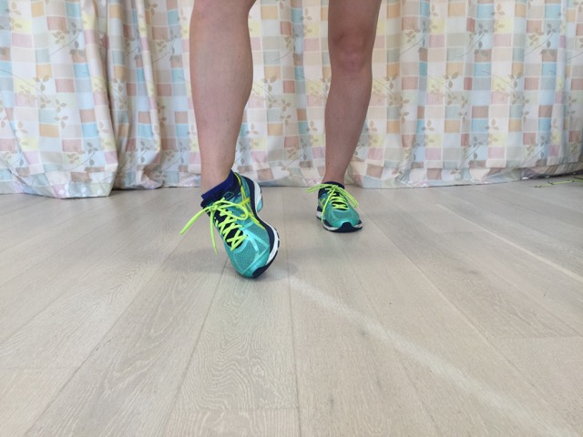
The usual position of an ankle sprain: inversion and some plantarflexion of the foot at the ankle joint. Permission: Joseph E. Muscolino.
Pain and swelling usually lessen as the ankle sprain becomes chronic. Most conditions are considered to be chronic once six months have passed since the initial injury. However, because it is difficult to avoid weight-bearing use of the injured side ankle, healing can take a very long time. For this reason, ankle inversion sprains, especially moderate or severe ones, sometimes require a year or more, for the pain and swelling to fully subside. Even once the overt pain is gone, the injured area often remains tender to touch.
Because the inversion ankle sprain results in stretched/torn lateral ligaments, chronic instability of the ankle joint usually results, and the injured ankle joint will display increased inversion range of motion permanently into the future.
Occasionally, if the ankle sprain is severe enough, pain and swelling may occur on the medial side of the ankle joint because the excessive inversion motion results in the bones on the medial side of the ankle jamming into each other. Further, due to the excessive inversion motion that occurs during the sprain, pain may also be present in the lateral leg due to strain of the eversion musculature (fibularis longus/brevis/tertius and extensor digitorum longus).
Assessment/Diagnosis of ankle sprain:
Assessment/diagnosis of an inversion ankle sprain of the ankle joint is based on verbal history as well as visual inspection, palpation, and range of motion (ROM) evaluation during the physical examination.
During verbal history, ask the client/patient to describe the position of the foot when the ankle sprain occurred; this can be helpful toward assessing which of the lateral ligaments was sprained. If the foot was sprained into pure inversion, the calcaneofibular ligament will most likely sustain the majority of the damage. Most commonly, because the client/patient is usually moving forward when the sprain occurs, the foot will also turn into plantarflexion along with the inversion and the anterior talofibular ligament will sustain the majority of the damage. Less commonly, if the foot is dorsiflexed as it was sprained into inversion, the posterior talofibular ligament will sustain the most injury.
The swelling will usually be apparent upon visual inspection. With Grade 2 and 3 sprains, ecchymosis (black and blue bruising) will often accompany the swelling. This occurs due to the pooled blood in the tissues that results from broken blood vessels internally. It is important to realize that pooled blood will usually descend with gravity, so the bruising will often show lower than the actual site of injury. If the swelling is not severe, it can be easily missed. So, for minor sprains, check for swelling by comparing the contour of the lateral malleolus on the injured side to the non-injured side; the visual clarity of the bony contour on the injured side is often diminished due to the swelling around the bone. Soft tissue swelling can also be detected by manual palpation assessment.
Palpation is also important for determining the location of pain to assess which ligament(s) is/are injured, as well as to have a sense of how severely damaged each ligament is. The sooner after the injury the palpation can be done, the better, because once the swelling forms, it can block the ability to palpate the underlying structures.
Passive ROM can also be done. Because inversion ankle sprains result in the tearing of laterally placed ligaments, the client’s/patient’s inversion passive ROM will usually be increased (the normal healthy foot should invert approximately 20 degrees from anatomic position). Assuming the client/patient has not sprained the other ankle, inversion ROM will likely be greater on the side of injury, so comparing left and rights can be helpful. It should be kept in mind that the presence of swelling can decrease the client’s/patient’s ROM, so an accurate evaluation of ankle ROM might have to wait until the swelling has subsided. Further, due to the tenderness of the region, if passive ROM is attempted, it should be done very gently.
It is also important to assess the eversion musculature because it might be overstretched and torn along with the ligament tissue. In other words, the client/patient might have experienced an ankle joint muscular strain along with the ligamentous sprain. Because the tissues on the medial side of the ankle joint are approximated during inversion, it is worthwhile to also assess the medial ankle joint region as well for compression injury.
It is not necessary to take X-Rays to assess/diagnose a ligamentous sprain. However, if the client/patient has experienced a moderate or severe sprain it is wise to have films to rule out an osseous fracture. A powerful sprain into inversion can possibly result in one of the lateral ligaments remaining intact and instead causing an avulsion fracture at its bony attachment, usually the lateral malleolus. It is also possible for the approximation of bones on the medial side to result in a crush fracture there as well.
Differential assessment of ankle sprain:
Whenever a client/patient has sustained a powerful inversion ankle sprain, the force that sprains the ligamentous tissue can easily damage other tissue. Therefore, it is important to also assess for other conditions. Chief amongst these is a strain of the eversion musculature and/or avulsion fracture on the lateral side of the ankle or possibly a crush fracture on the medial side. If the force is powerful enough, it is also possible to dislocate the ankle joint, subtalar joint, or even the transverse tarsal joint (located between the talus and calcaneus proximally and the navicular and cuboid distally; technically composed of the talonavicular joint and the calcaneocuboid joint).
