Learn how growth hormone affects muscle recovery, collagen synthesis, lean mass and strength, and what research says about medical HGH treatment.
Learn how growth hormone affects muscle recovery, collagen synthesis, lean mass and strength, and what research says about medical HGH treatment.
Being able to feel tissue tension barrier is the one most crucial aspect for a clinical orthopedic manual therapist performing deep pressure massage. From a mechanical standpoint, it is only pressure beyond the tissue tension barrier that effects therapeutic change (this is not necessarily true neurally).
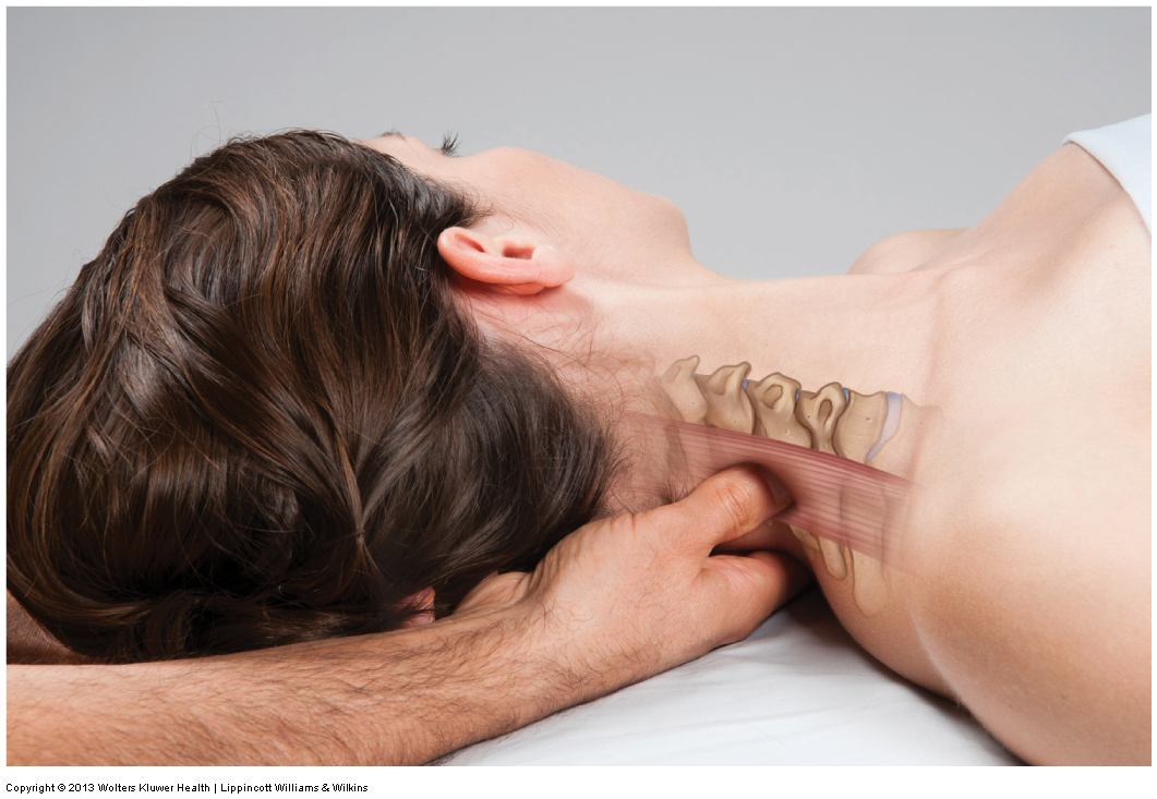
When performing deep pressure massage, deep stroking massage must originate from your core by further rocking your pelvis and extending your spine forward. Short deep strokes to the neck between 1 and 2 inches (2-5 centimeters) in length allow you to preserve optimal body mechanics.

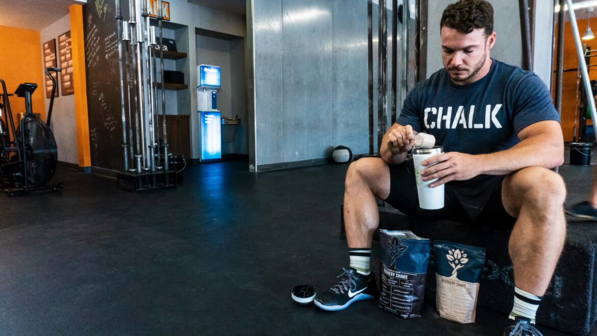
The moment you start selling a physical product, you run into a problem most therapists never trained for: taking a product photo good enough to actually sell something.

Somatic therapy addresses what clinical care often misses by meeting you in your nervous system. It’s not meant to replace clinical work. It’s there to complement it.

That people aged 18-21 in this study have not yet begun to experience pain as a result of text neck posture does not surprise me at all. They are simply still too young to experience what the physical stress of the overuse of text neck posture will eventually do to them.
In summary, exercise-based rehabilitation is the best treatment for tendon pain. A progressive program that starts with a strength program and then progresses through to more spring-like exercises, and endurance aspects will give the right loads on the tendon and the best long-term results.

This article is provided as general professional information. It is not intended to be used for individual treatment of a client who has had an ACL reconstruction.

Health problems can range from the ones that are minor to those that can be pretty serious, even if they aren’t life-threatening. And that’s something that can take a toll on a person’s mental health.
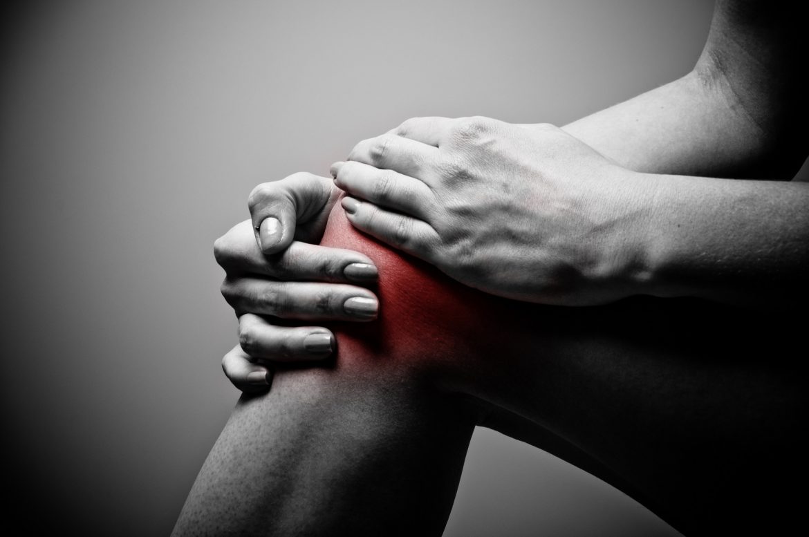
Once one joint/structure/tissue is injured, compensation patterns usually occur in which the individual offloads physical stress to other areas of the body. This would logically lead to an increased risk of use/overuse/misuse/abuse to these other areas, likely eventually leading to injury.
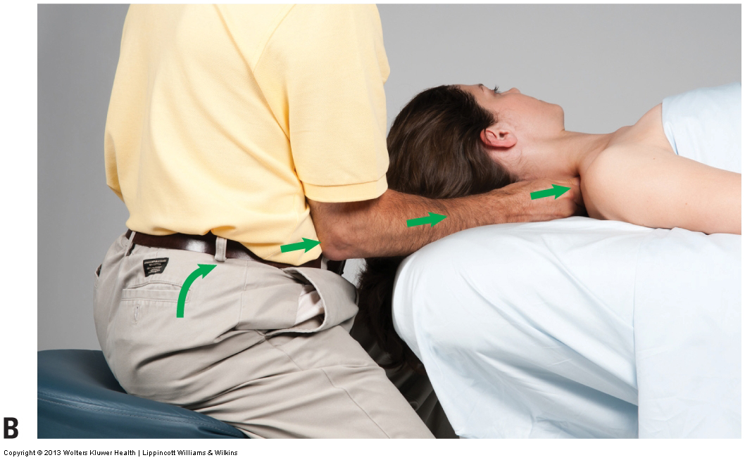
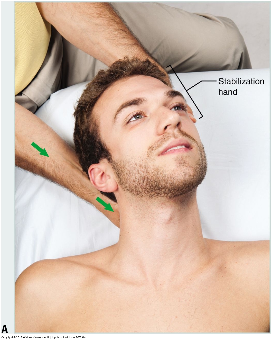
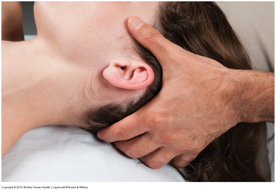
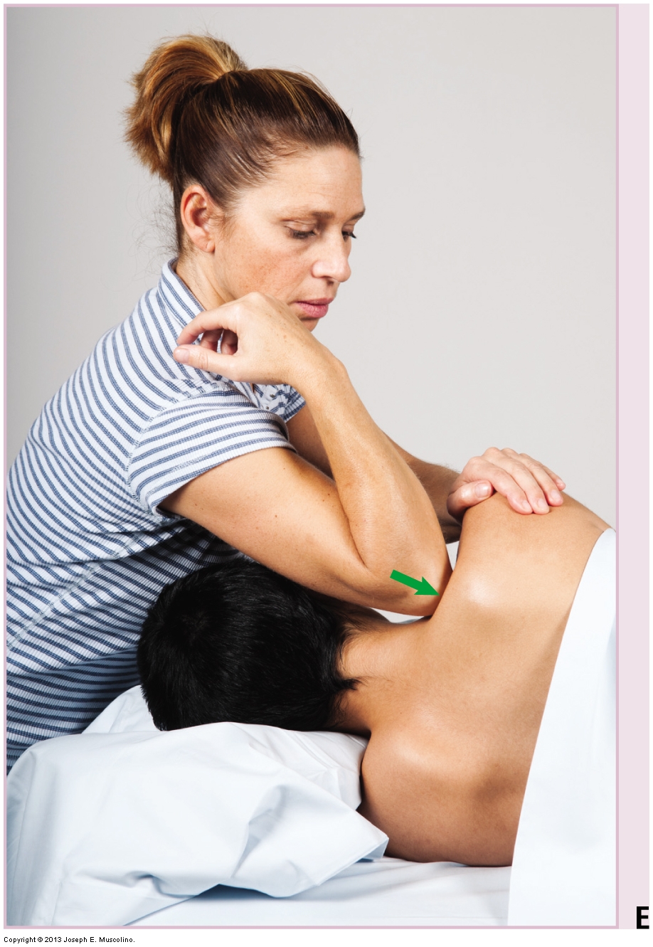
For deep pressure massage to the neck, align your core with your stroke by laterally (externally) rotating your arm at the shoulder joint so that your elbow is positioned in front of your core. Now lock your elbow into your core just inside (and usually slight above) your anterior superior iliac spine.
The five programs here vary considerably. Two take a university-led approach to nutrition science and behaviour change. The others offer shorter routes for professionals who mainly want practical tools for client conversations.

If you’re a registered nurse, you already know the job asks a lot from your brain, your feet, and sometimes your patience before breakfast. It’s rewarding, but it can also leave you wondering what comes next.
When performing deep pressure massage into the neck, maximal pressure for minimal effort is achieved if the angle of your pressure into the client is perpendicular to the contour of the neck where you are working.
Even perfect body mechanics cannot eliminate all physical stress to your body when doing massage. Ideal body mechanics merely minimize the stress. For this reason, when doing massage, especially deep pressure massage, it is wise to alternate which treatment hand contact you use during a session.

Yes, chronic disease has changed what healthcare looks like. Care doesn’t begin when you walk into a clinic. It doesn’t end when you leave. It’s part of everyday life.

Chronic TMJ problems can repeat themselves. The early evaluation and treatment for your jaw pain can help you find the best ways to manage your symptoms and to protect your jaw from future damage.
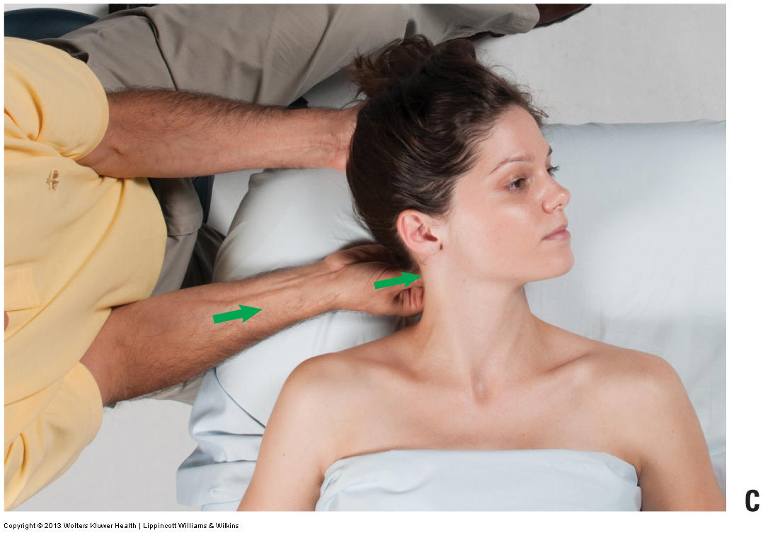
When performing deep pressure massage for the neck with the client supine, it is typical for the therapist to sit, centered at the head of the table. However, this does not allow for efficient body mechanics because it is difficult or impossible to position your core in line with the stroke.
The science of performing deep tissue work to the neck follows the laws of physics and, whenever possible, involves the use of body weight and the contraction of larger muscles instead of smaller ones. The art of performing deep tissue work lies in exactly how these guidelines are carried out and applied.

IV therapy earns its place when there is a measurable deficit to correct and a genuine reason oral intake will not do the job.
Here’s an honest walk-through of what becoming a Pilates instructor online actually involves in 2026 — the steps, the timelines, the costs and the things worth checking before you enrol.
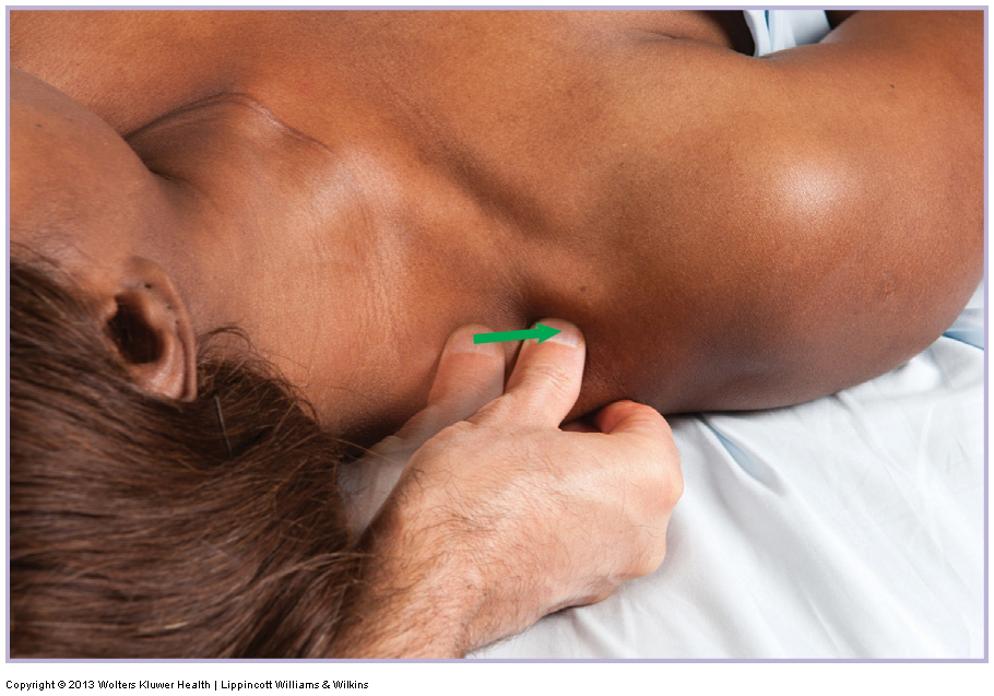
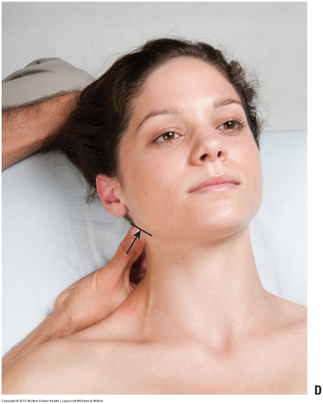
Side-lying position for deep pressure massage into the neck can be very effective and can allow for effective longitudinal as well as transverse cross-fiber strokes; however, it is important to avoid exerting deep pressure too far anteriorly onto the transverse processes of the client’s neck.
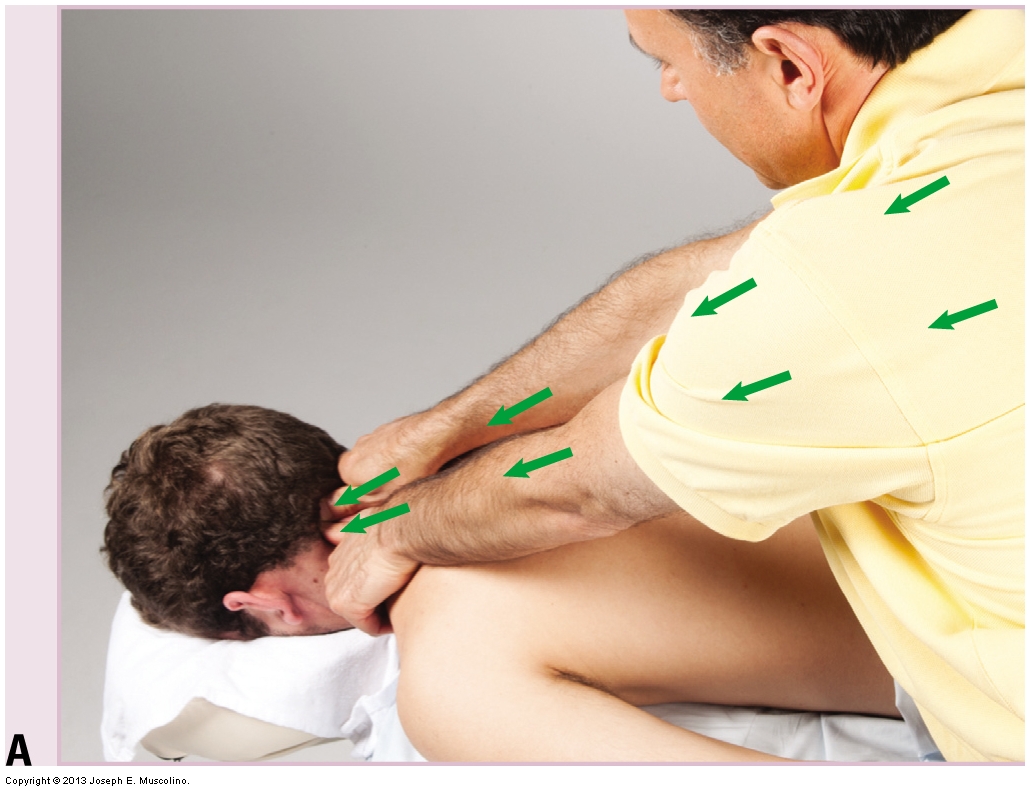
We can take advantage of our body weight to generate deep pressure massage into the client’s tissues by simply leaning into the client. Pressure derived this way is effectively free because it takes no effort on our part. For this reason, it should be used whenever possible.
The truth is, back problems rarely come out of nowhere. They build quietly. Tiny habits. Tiny shortcuts. Tiny moments repeated hundreds of times until your body finally decides it’s had enough.
For aspiring nursing assistants, the takeaway is straightforward: understanding body systems strengthens every part of the role.
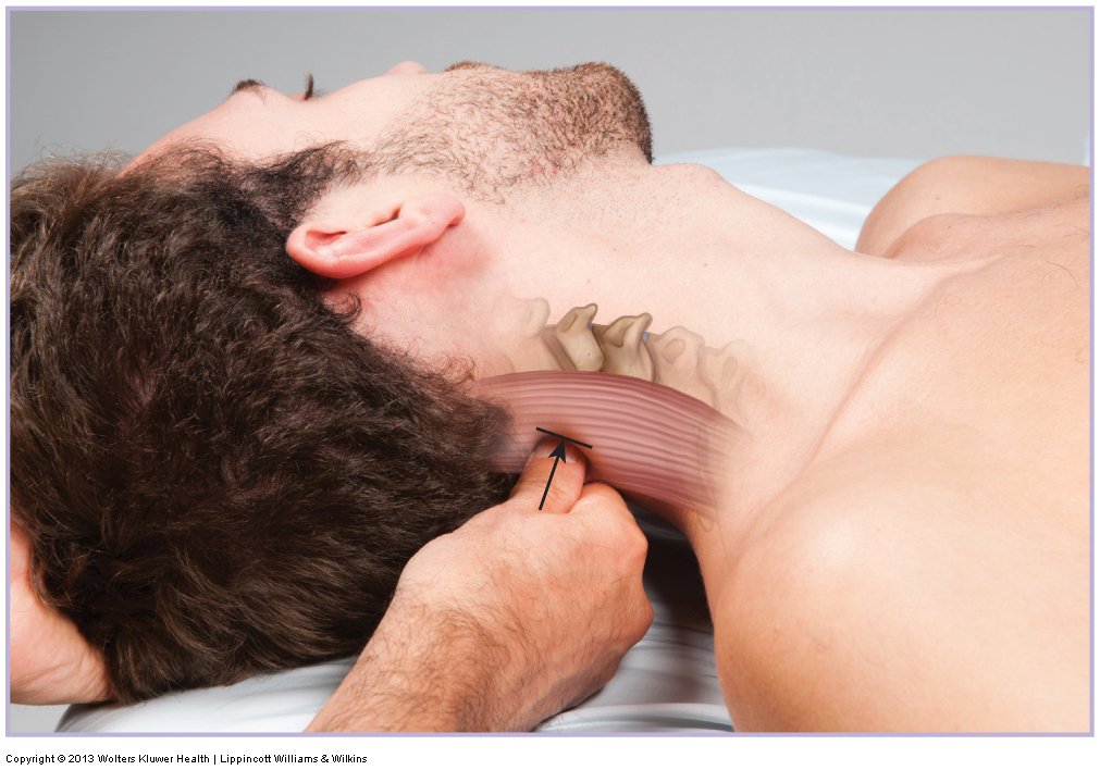
Force for deep pressure can be generated in two ways: externally or internally. The external generation of force comes from the force of gravity by using our body weight. The internal generation of force comes from the contraction of our muscles.

Running economy for distance runners is a complex multi-factorial measure of running efficiency, which reflects the combined functioning of biomechanical, neuromuscular, metabolic, and cardio-respiratory factors, some of which are hereditary and some of which adapt to coaching.

Laser hair removal can simplify your grooming routine, reduce skin irritation, and help you spend less time worrying about shaving and more time doing the activities you enjoy. The only question is, is it the right choice for you?
This article explores the point at which hands-on treatment stops being sufficient on its own, and the foot complaint is better co-managed with a podiatrist.
The therapist performed a static assessment of the client’s posture and noted the typical upper crossed syndrome with a hyperkyphotic thoracic spine, a hypolordotic lower cervical spine, a hyperlordotic upper cervical spine, protracted head, protracted scapulae, and medially (internally) rotated arms.